Download

1 / 17
170 likes | 186 Views
Learn about the intricate structures of the eye orbit and eyelids, nerve innervations, and potential clinical implications. Explore the bony orbit, optic nerves, lacrimal glands, and more. Essential knowledge for medical education.

E N D
DEMO-VI(Eye) Ali JassimAlhashli Year IV – Unit VIII - CNS
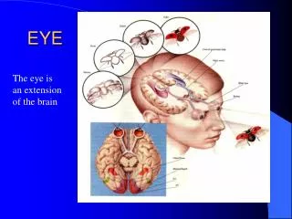
STATION-1 • The bony orbit: it is protecting our eyeball and resembling a pyramid: • With a base directed: anterolaterally. • And an apex directed: posteromedially. • Notes: • The medial wall of the pyramid is extending farther anteriorly than the lateral wall. • The orbital axes diverge at 45 degrees while the optical axes are parallel to each other.
STATION-1 • The walls of the orbit are formed by the following bones: • Roof of the orbit: frontal bone (mostly) + lesser wing of sphenoid bone. • Floor of the orbit: maxillary bone + zygomatic bone (meeting at the zygomato-maxillary joint). • Lateral wall of the orbit: frontal bone + zygomatic bone (mostly) + greater wing of sphenoid bone. • Medial wall of the orbit: sphenoid body + ethmoid bone + lacrimal bone + maxillary bone.
STATION-1 • In the image of previous slide: • Supraorbital notch: is for the passage of supraorbital nerve which is branch of ophthalmic division of trigeminal nerve. Notice that this nerve is important to elicit deep pain. • Infraorbital foramen: for the passage of infraorbital nerve which is a branch of maxillary division of trigeminal nerve. • Lesser wing of sphenoid bone contains the optic canal through which the optic nerve (2nd cranial nerve) and ophthalmic artery pass. • Lacrimal bone contains the nasolacrimal duct which is involved in the drainage of tears to the nasopharynx. • Notice that the medial wall of the orbit is very thin. • Structures passing through the superior orbital fissure are: • Opthalmic veins. • Ophthalmic division of trigeminal nerve. • 3rd, 4th and 6th cranial nerves. • Structures passing through the inferior orbital fissure are: • Infraorbital vessels. • Inferior division of ophthalmic vein.
STATION-1 • Medial and lateral palpebral tendons of the eye are extensions of orbicularisocculi muscle (the circular muscle which is surrounding the eye). • Tunica bulbi: it is enclosing the eyeball and allowing its rotation. Check ligaments are extensions from it.
STATION-2 • Lacrimal glands are in the superolateral aspect of the eye. They are composed mainly of 2 parts: • Deep orbital lobe. • Superficial palpebral lobe. • Lacrimal glands are innervated by the facial nerve and the lacrimal nerve which is a branch of the ophthalmic division of trigeminal nerve.
STATION-2 • Nerves of the orbit: • Optic nerve (2nd cranial nerve): entering the orbit from middle cranial fossa through the optic canal (accompanied by ophthalmic artery). • Ophthalmic division of trigeminal nerve will give 3 branches • Lacrimal nerve: enters the orbit through the superior orbital fissure. • Frontal nerve: divides into supratrochlear and supraorbital nerves. • Nasociliary. • Oculomotor nerve (3rd cranial nerve): entering the orbit through superior orbital fissure. • Trochlear nerve (4th cranial nerve): entering the orbit through superior orbital fissure. • Abducens nerve (6th cranial nerve): entering the orbit through the superior orbital fissure.
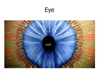
STATION-2 • Anatomy of the eyelid: • Eyelids are movable folds which cover the eyeball anteriorly. • It is covered externally by skin and internally by a mucous membrane called conjunctiva (notice that conjunctiva does not cover the cornea). • The structures which are strengthening the eyelid superiorly and inferiorly are known as superior and inferior tarsi. • Eye lashes are attached to the margins of the eyelid and they are connected with sebaceous glands. • Tarsal glands: they secrete lipids which lubricate edges of the eyelids and prevent them from sticking when they are closed.
STATION-2 • Ciliary ganglion: parasympathetic ganglion in the posterior part of the orbit between the optic nerve and lateral rectus. • Ophthalmic artery (which is passing through the optic canal) is a branch of internal carotid artery. It will give the following branch: central retinal artery (which is an end artery and any occlusion in it can result in blindness ).
STATION-2 • Superior ophthalmic vein is passing in the superior orbital fissure. • Inferior ophthalmic vein is passing in inferior orbital fissure. • Injury to the suspensory ligament of the eye → eyeball will fall → resulting in diplopia.
STATION-3 • General movements of the eye:
STATION-4 • Sympathetic nervous system is innervating: • Sweat glands → this explains the anhidrosis. • Palpebral fissure → therefore, when injured → it will result in partial ptosis. • Sympathetic nervous system is from T1 – (L2/L3). • Parasympathetic nervous system (craniosacrum): • Cranial part from: 3,7,9 and 10. • Saral part from: S2, S3 and S4. • Sympathetic chain/ trunk: • Upper thoracic part: is responsible for innervating the heart and lungs. • T5- (L1/L2) part: is represented by the splanchnic nerves (greater, lesser, least) which are synapsing in prevertebral ganglions: • Celiac. • Superior mesenteric. • Inferior mesenteric. Note: a part of the greater splanchnic nerve which is not synapsing is the one which is going directly to adrenal medulla.
STATION-4 • Parasympathetic nervous system: • From nuclei of cranial nerves to the following ganglia: • Submandibular: innervating the mandible and submandibular salivary gland. • Otic: innervating parotid cerebri gland. • Pterygopalatine: innervating lacrimal gland. • Ciliary: going to Edenger-Westphal nuclei and innervating ciliary muscle and constrictor papillae. • Parasympathetic nerve fibers from S2-S4 are forming the: pelvic splanchnic nerve (which is innervating the organs of the pelvis). • Innervation of internal muscles of the eye: • Sympathetic nerve fibers: innervating radial muscle of the iris (which causes dilation of the pupil). • Parasympathetic nerve fibers: innervating circular muscle of the iris (which causes constriction of the pupil) and ciliary muscle.