Download
1 / 59
590 likes | 744 Views
Safe Working With Ionising Radiation Revised January 2012. John Sutherland, University Safety and Radiation Protection Officer. Remember . Please make sure you have signed in - otherwise you will need to re-attend!! Handout - also downloadable from Safety Office Web Page. Programme.

E N D
Safe Working With Ionising RadiationRevised January 2012 John Sutherland, University Safety and Radiation Protection Officer
Remember • Please make sure you have signed in - otherwise you will need to re-attend!! • Handout - also downloadable from Safety Office Web Page
Programme • What is radiation? • How is it measured? • Biological harm • Doses into perspective • Legislation • Unsealed work • X-ray/Sealed - Harry Zuranski, Safety Office.
Objectives • Foundation for Training in School • Understand principles • radiation types and effects • biological effects • relative risk • legislation • university arrangements • Safe Practice
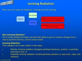
Isotopes • Variable neutron number • Unstable nuclei transform • Ionising radiation emitted
Ionisation • Energy transfer • Enough energy ~ 13+ eV
Half - life Isotope Half-Life Tritium 12.4 y Carbon 14 5730 y Sulphur 35 87.4 d Phosphorus 33 25.6 d Phosphorus 32 14.3 d Iodine 125 60.1 d
Types of Radiation • Video
Types of Radiation • Alpha • From heavy nuclei (e.g. Americium 241) • Helium nuclei (2P+2N) • 1500 ionisations • Dangerous internally • Easily shielded as very large particles • Sheet of paper or plastic film • Small distance of air • Dead outer layer of skin
Types of Radiation • Beta Particles (B) • High speed electrons from nucleus • Identical to orbital electrons • Neutron Proton + B- • Energy dependent penetrating power • 3H - 18.6 KeV • 14C - 156 KeV • 32P - 1.71 MeV • Rule of thumb for maximum range of beta particles • 4 metres in air per MeV of charge • P32 can travel up to 7 m in air but 3H only 6mm! • Easily shielded with perspex, higher energy needs greater thickness • 10 mm will absorb all P32 betas • Cannot reach internal organs
Types of Radiation • Bremsstrahlung • X-radiation resulting from high energy ß particle absorption in high density shielding, e.g. lead. • Risk with 32P and similar high energy ß emitters. • Shield ß with lightweight materials such as perspex. • Very large activities can still produce some Bremsstrahlung from perspex - supplement perspex with lead on outside to absorb the X-rays.
Types of Radiation • Gamma Radiation (Y) • Electromagnetic radiation • Emitted from nucleus • Readjustment of energy in nucleus following a or ß emission • Variable energy characteristic of isotope • Highly penetrating • 5 - 25 cm lead • 3m concrete • Can reach internal organs • Can pass through the body
Types of Radiation • X-Radiation • Similar to gamma but usually less energetic • Originates from electron cloud of the nucleus • Produced by machines - can be switched off! • Also produced by some isotopes • Iodine-125 produces both gamma and x-rays • Broad spectrum of energy
Types of Radiation • X-rays • Incident radiation ejects electron • Outer electron fills gap • X-ray energy = difference between orbital energy levels - characteristic • Bremsstrahlung also produced
Types of Radiation • Neutrons • Large, uncharged, physical interaction. • Spontaneous fission (Californium 252) • Alpha interaction with Beryllium (Am-241/Be) • Shield with proton-rich materials such as hydrocarbon wax and polypropylene. • Americium/Beryllium sources are used in neutron probes for moisture or density measurement in soils and road surfaces etc. These also emit gamma radiation.
Units of Radiation • SI units Becquerel, Gray, Seivert • replaced Curies, Rems, Rads • Activity • Dose • absorbed • equivalent • committed
Units of Radiation - activity • Quantity of r/a material • Bequerel (Bq; kBq; MBq) • 1 nuclear transformation/second • 3.7 x 10¹⁰ Bq = 1 Curie • Record keeping • Stock, disposals • Expt protocols
Units of Radiation - dose • Absorbed - Gray (Gy) • Radiation energy deposited • 1 Gy = 1 joule/kg • Dose Equivalent - Seivert (Sv) • modified for relative biological effectiveness • beta, gamma, X = 1 • alpha, neutrons = 10-20
Units of Radiation - committed • Internal • irradiation until decay or elimination • radiological and biological half-lives • data for 50-year effect • Annual Limit on Intake (ALI) • limit on committed dose equivalent • quantity causing dose limit exposure
Exposure to Ionising Radiation • Environment • Naturally occurring radioactive minerals remaining from the very early formation of the planet. • Outer space and passes through the atmosphere of the planet – so-called cosmic radiation. • Man-made • medical treatment and diagnosis. • industry, primarily for measurement purposes and for producing electricity. • fallout from previous nuclear weapon explosions and other accidents/incidents world-wide.
Biological Effects of Radiation Exposure • Ionising radiation affects the cells of the body through damage to DNA by: • Direct interaction with DNA, or • Through ionisation of water molecules etc producing free radicals which then damage the DNA. • Some damaged cells might be killed outright so do not pass on any defect. • In some cases cell repair mechanisms can correct damage depending on dose.
Biological Effects of Radiation Exposure • Deterministic Effects. • Threshold beneath which there is no effect and above which severity increases with exposure. • High dose effects - cells may be killed by damage to DNA and cell structures. • Clinically observable effects include: • 5 Sv to whole body in a short time is fatal. • 60 Sv to skin causes irreversible burning. • 5 Sv to scalp causes hair loss • 4 Sv to skin causes brief reddening after three weeks • 3 Sv is threshold for skin effects.
Biological Effects of Radiation Exposure • Stochastic (Chance) Effects • No threshold dose, probability of effect increases with dose but severity of effect remains unchanged • Lower dose effects • No obvious injury, • Some cells have incorrectly repaired the DNA damage and carry mutations leading to increased risk of cancer. • Rapidly dividing cells most at risk – blood forming cells in bone marrow; gut lining.
Main Area of Available Data for Study E F F E C T Main Area of Interest for Radiation Protection RADIATION DOSE Cancer Risk at Low Doses • Evaluation of Cancer Risk • Studied for decades. • atomic bomb explosions in Japan, • fallout from nuclear weapons tests • radiation accidents. • medical irradiations, • work (e.g. nuclear power industry) • living in a region that has unusually high levels of radioactive radon gas or gamma radiation.
Cancer Risk at Low Doses • Life-time risk of cancer from all causes of about 20–25%. • Exposure to all sources of ionising radiation (natural plus man-made) could be responsible for an additional risk of fatal cancer of about 1% • Dose from natural background radiation is about 2.2 mSv per year. • Dose from non-medical, man-made radiation • 0.02 to 0.03 mSv per year (1/100th natural background), • 0.01% of additional cancer risk. • More significant cancer risk factors include: • cigarette smoking, • excessive exposure to sunlight, and • poor diet.
Biological Effects • 4-10 Sv - death • 1 Sv - clinical effects • 100 mSv - clinical effects on foetus • 50 mSv - max lifetime univ. dose • 20 mSv - annual whole body dose limit • 6 mSv - classified worker • 2.5 mSv - average annual exposure (UK) • 1 mSv - foetus after pregnancy confirmed • 150 - 250 uSv - max annual dose at univ. • 20 uSv – average annual dose at univ.
Perspective on Exposures • Nature of work AND precautions in place show risk from exposure at work is extremely low. • 10-15% of those subject to dosimetry receive a measurable dose, • Average dose ~ 18uSv • 0.1% of the dose limit of 20 mSv, • 1% of that received from natural background radiation (2.2 mSv). Follow Safe Procedures
Legislation • Health and Safety • Ionising Radiations Regulations 1999 • Environmental • Environmental Permitting Regulations 2010 • (Supersede Radioactive Substances Act 1993)
Ionising Radiations Regulations 1999 • Worker protection • dose limits • Justification • Radiation Project Proposal Forms (Rad 1-3) • risk assessment for exposure • Risk Assessment Forms (Rad 5 or 6) • restrict exposure through • equipment, procedure, experimental design • time, • shielding, • distance (inverse square law)
Protection through distance • Inverse square law applies Distance Dose rate (uSv/hr) 1m1 2m 0.25 4m0.06
Protection through distance • HOWEVER !!!!!! • Distance Dose rate (uSv/hr) • 100cm 1 • 50cm 4 • 30cm 9 • 10cm 100 • 1cm 10,000 • 1mm 1,000,000
Ionising Radiations Regulations 1999 • Local Rules • RPS’s for all areas • Worker/Project registration • Designation of areas • access control • contamination monitoring • Worker responsibility • Regular checks by RPS • Secure storage and accounting • Movement • packaging and labelling • No posting or carriage on public transport
Environmental Permitting Regulations 2010 • Enforced by Environment Agency. • Licensing regime • stocks • accumulation and disposal of waste • specific limits on • isotope and quantity, • disposal route and disposal period • Strict record keeping essential • Isostock for Radiochemicals • Must be kept up to date
Administrative Controls • Project Registration (Rad 1-3) • Isotopes • Quantities • Disposal routes • Lab Facilities • Worker Registration (Form) • Project • Dosemeter • Look after it • Return at end of quarter – charges for late/lost badges • Amend Details if Work Changes
The Use of Radiochemicals in Life Science Research Comparison of Common Isotopes Safe Handling – 10 Golden Rules Decomposition
Carbon-14 • Low energy b emission - no shielding required • Long half-life - less time pressure • Low specific activity - low sensitivity • Detection • scintillation counter • autoradiography • Geiger counter • phosphorimager • Labelled compounds generally stable - few decomposition problems
H-3 (Tritium) • Very low energy b emission - no shieldingrequired • Long half - life • High specific activity - reasonably sensitive, but weak emission • Detected by • scintillation counter detection less easy • autoradiography less accurate and • fluorography less efficient than 14C • phosphorimager • Labelled compounds less stable - radiation decomposition problems
Iodine -125 • g emission - lead shielding required • Short half-life - time pressures • Very high specific activities - high sensitivities • Detection • Gamma counter • Scintillation probe • Autoradiography • phosphorimager • Labelled compounds stable - some decomposition problems
Phosphorus - 32 • High energy b emission - shielding required (perspex and lead) • 1 MBq in 1ml plastic vial @ 1m 2.5uSv/hr @ 10cm 200uSv/hr • 30MBq in 1ml plastic vial @ 10cm 6mSv/hr • 25 hours of work = 150mSv, i.e.Classified Worker NEVER HOLD VIAL IN FINGERS
Phosphorus - 32 • High energy b emission - shielding required (perspex and lead) • Short half-life - time pressures • Very high specific activity - very high sensitivity • Detection • Scintillation counter • Cerenkov counter • Geiger counter • Autoradiography • phosphorimager • Labelled compounds unstable - decomposition problems
Phosphorus - 33 • Low energy b emission - low shielding required (1cm perspex) • Short half -life - time pressures • High specific activity - high sensitivity • Detection • Scintillation counter Easy to detect • Proportional counter and accurate counting • Geiger counter • Autoradiography • phosphorimager • Labelled compounds generally stable - few decomposition problems
Sulphur -35 • Low energy b emission - low shielding required (1cm perspex) • Shortish half-life - some time pressures • High specific activity - high sensitivity • Detection • Scintillation counter • Proportional counter • Geiger counter • Autoradiography • phosphorimager • Labelled compounds generally stable - few decomposition problems
Resolution Intensifying screen Plastic base aasAS Emulsion Anti scratch H-3 C-14/ S-35/ P-33 P-32/ I-125 Image on film: Blank
Choosing an isotope • Detection method • Resolution required • Sensitivity • Specific activity • Formulation - aqueous/ethanol (stabilised/free radical scavenging) • Position of label - important in metabolic studies / can affect protein binding
Working safely with radioactivity The Ten Golden Rules • Understand the nature of the hazard and get practical training • Plan ahead to minimise handling time • Distance yourself appropriately from sources of radiation • Use appropriate shielding • Contain radioactive materials in a defined work area • Wear appropriate protective clothing and dosimeters • Monitor the work area frequently • Follow the local rules and safe ways of working • Minimise accumulation of waste and dispose of it correctly • After completion of work monitor yourself and work area
Decomposition • Chemical decomposition caused by, or accelerated by: • the presence of one or more radioactive atoms in the molecule • Free radicals • Micro-organisms • Stock solutions and aliquots will decompose over time and become unusable.