Download
1 / 44
440 likes | 1.14k Views
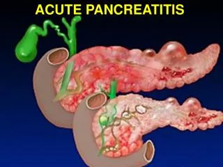
Introduction . Acute pancreatitis is an inflammation of the pancreas. Associated with edema, pancreatic autodigestion, necrosis and possible hemorrhage. Acute pancreatitis (AP) is a common disease that normally runs a benign course in the majority of patients. However in up to 20% of individuals th

E N D
1. Management of Acute Pancreatitis
MR ??? MA ???
2. Introduction Acute pancreatitis is an inflammation of the pancreas. Associated with edema, pancreatic autodigestion, necrosis and possible hemorrhage.
Acute pancreatitis (AP) is a common disease that normally runs a benign course in the majority of patients. However in up to 20% of individuals the disease is severe and may be associated with a mortality close to 20%.1,2
3. Terminology and definitions The 1992 Atlanta International Symposium on Acute Pancreatitis put forth a classification system of acute pancreatitis with the intent of providing clinical guidance and an exact vocabulary across institutions and within the literature. 3
4. Epidemiology of acute pancreatitis Estimated incidences range from 5.4/100 000 population per year in England to 79.8/100 000 in the USA. 4,5
there appears to be an increase in the incidence of acute pancreatitis.6
This rise in incidence has been attributed to increased alcohol consumption in Finland and the Netherlands, but may well reflect improved diagnostic capability during this period.
No seasonal or weekly pattern of acute pancreatitis has been observed.7
Men are affected much more than women and the main age group affected is 40�60 year olds.8
5. Etiology of acute pancreatitis Gallstones: Gallstones continue to be the leading cause of acute pancreatitis in most series (30�60%). Microlithiasis (occult gallstones) is a well-known cause of acute pancreatitis. Biliary microscopy and endosonography are the recommended tests to diagnose microlithiasis.9,10 Many studies have shown that microlithiasis is the cause of presumed idiopathic acute pancreatitis in 50�73% of patients. 11,12,13
6. Alcohol: It is responsible for about 30% of all cases of acute pancreatitis. The proponents of the necrosis-fibrosis hypothesis, indeed, believe that repeated attacks of acute alcoholic pancreatitis lead to chronic pancreatitis.14
Hyperlipidemia: Hyperlipidemia generally associated with serum triglyceride levels > 1000 mg/dL is the cause of acute pancreatitis in about 1.3�3.8% of cases. Patients with diabetes or those on certain drugs may have high triglyceride levels causing pancreatitis.
Idiopathic acute pancreatitis: About 10% of patients are left with no identifiable cause despite a thorough biochemical, ultrasonographic and endoscopic examination.15,16
7. Clinical presentation The cardinal symptom of acute pancreatitis is moderate to severe epigastric pain.
The pain radiates to the back quite commonly owing to the retroperitoneal location of the pancreas. It may also radiate to the flanks, chest, shoulders and lower abdomen.
The character of the pain is steady and boring, but not colicky.
Nausea and vomiting are other common symptoms of acute pancreatitis.
Fever in the first week is due to acute inflammation and is mediated by inflammatory cytokines. Fever in the second or third week in patients with acute necrotizing pancreatitis is usually due to infection of the necrotic tissue and is much more significant.
8. The infection in patients with acute pancreatitis is usually due to Gram-negative bacteria (E-coli, enterobacter).
Cardiopulmonary Exam
Tachycardia, Hypotension: hypovolemia or vasodilation and initial systemic inflammatory response syndrome (SIRS)
Hypoxemia (25%)
Left basilar rales (Pleural Effusion)
9. Abdominal Exam
Abdominal tenderness and rigidity
Bowel sounds decreased
Palpable upper abdominal mass Acute fluid collections and pseudocysts
Cullen's Sign (periumbilical discoloration)
Turner's Sign (flank discoloration) * blue-gray discoloration of abdominal flanks due to exudation of blood-stained fluid into the subcutaneous tissue, usually 72 h into the illness.
Skin Exam
Erythematous skin Nodules (Subcutaneous Fat Necrosis)
11. Diagnosis: Biochemical Serum Amylase elevated
Nonspecific
Returns to normal in 48-72 hours
Normal amylase does not exclude pancreatitis
Level of elevation does not predict disease severity
Serum Lipase elevated
Specific for pancreatic disease
Returns to normal in 7-14 days
Serum Electrolytes
Hypocalcemia (25%)
Hyperglycemia Complete Blood Count (CBC)
White Blood Cells increased to 15k-20k
Lipids Elevated
Hypertriglyceridemia (15%)
Liver Function Tests
Serum Bilirubin elevated
Alkaline Phosphatase elevated
Aspartate Aminotransferase elevated (AST)
Hypoalbuminemia (Poor prognosis)
Lactate Dehydrogenase (LDH) elevated (Poor prognosis)
12. Severity assessment The presence of a high APACHE II SCORE at admission (8 or greater), a pleural effusion, a high body mass index, evidence of necrosis on contrast-enhanced CT (CECT) and a CRP level greater than 150 mg/L at 48 h are all useful markers of severe disease.
Infection of the necrotic tissue after the first week of illness is the major determinant of later outcome. However, it is organ failure in which respiratory failure dominates that determines outcome in the majority of difficult cases to manage.
14. Ranson's criteria On admission�
Age > 55 yrs
WCC > 16,000
LDH > 600 U/l
AST >120 U/l
Glucose > 10 mmol/l Within 48 hours
Haematocrit fall >10%
Urea rise >0.9 mmol/l
Calcium < 2 mmol
pO2 < 60 mmHg
Base deficit > 4
Fluid sequestration > 6L
15. Severity assessment Glasgow scoring system for the prediction of severity in acute pancreatitis
16. amylase clearance Amylase clearance is used to differentiate between a patient with macroamylasemia secondary to hyperamylasemia from a patient with pancreatitis
normal ratio for amylase clearance is between 2-5%
in pancreatitis, the ratio is increased
amylase clearance= ( urine amylase concentration ) x (serum creatinine concentration )/ ( serum amylase concentration ) x ( urine creatinine concentration )
17. The role of imaging in the diagnosis and staging of acute pancreatitis Dynamic contrast-enhanced CT (CECT) is the imaging modality of choice for diagnosis, staging, and detection of complications of acute pancreatitis. Other techniques, such as ultrasonography, ERCP, and angiography are useful for problem solving and for specific evaluation of the pancreatic and biliary ducts and vascular system .
Most importantly, CECT has been shown to have a sensitivity of 87% and an overall detection rate of over 90% for pancreatic gland necrosis.18,19,20
18. Balthazar
19. GradeCriteria:
A: Normal
B: Focal or diffuse glandular enlargement, Small intra-pancreatic fluid collection
C: Any of the above, Peripancreatic inflammatory changesLess than 25% gland necrosis
D: Any of the above, Single extrapancreatic fluid collection25-50% gland necrosis
E: Any of the aboveExtensive extrapancreatic fluid collectionPancreatic abscessMore than 50% gland necrosis
20. CT Severity index serial CT scans are important for following the progression of the disease and for detecting additional complications.
In Balthazar�s series
21. Acute pseudocyst
22. Pancreatic necrosis
23. Peripancreatic and retroperitoneal edema
24. Immediate assessment Clinical assessment including great care to assess respiratory, cardiovascular and renal compromise.
Body mass index. There is considerable risk (> 30 kg/m2) or much greater risk > 40 kg/m2
Chest X-ray. Is there a pleural effusion present?
Contrast-enhanced CT. Is there more than 30% of the volume of the pancreas malperfused?
APACHE II score. Is it 8 or greater?
Presence of organ failure.
25. Resuscitation Transudation of fluid from the intravascular space to the peritoneum is the principle cause of hypovolemia in AP.
Assessment of the patient�s volume status determined by heart rate, blood pressure, urine output and jugular venous pressure.
An infusion rate should be set that accounts for basal fluid requirements (35 mL/kg per day) and ongoing third space losses.
26. Potassium chloride should be added to the intravenous fluids to achieve 100 mEq/day.
Glucose levels greater than 13.9 mmol/L (250 mg/dL) necessitate insulin administration.
A blood transfusion is indicated if the patient�s hematocrit is less than 25%; values ranging from 30 to 35% are considered optimal for pancreatic parenchymal perfusion.21
27. Any evidence of respiratory insufficiency requires a chest X-ray to assess for pulmonary edema or acute respiratory distress syndrome (ARDS)
The use of H2 antagonists or proton pump inhibitors can ameliorate the tendency to metabolic alkalosis and prevent stress ulcers.22,23,24
28. Analgesia Patient comfort is essential. Patients with pain due to pancreatitis tend to have a high respiratory rate form hypoxic �drive�, which increases insensible fluid losses, decreased lung volumes from �splinting� and reduced mobilization, which hampers lung function and increases the risk of deep venous thrombosis.
Severe pain should be treated with meperidine 50 to 100 mg IM q 3 to 4 h prn in patients with normal renal function (morphine causes the sphincter of Oddi to contract and should be avoided).
29. Antibiotic prophylaxis Infectious complications are still regarded as the primary cause of mortality in severe pancreatitis.25 Thus, it is essential to identify the presence of pancreatic necrosis and take measures to prevent infection.
The current recommendation is the use of a systemic antibiotic such as imipenem-cilastatin 500 mg three times a day for 2 weeks 26,27,28 in patients with documented pancreatic necrosis.
30. Antibiotic prophylaxis An acceptable strategy would be to perform a CT scan with intravenous contrast at days 4�7 and begin imipenem if necrosis is present. The use of early antibiotic treatment with imipenem has been shown to decrease the need for surgical intervention.29
If there is clinical evidence of infection, pancreatic necrosis should be sampled by CT-guided fine needle aspiration (FNA).30 If infection is confirmed, the tissue should be treated by surgical debridement, either via open access or percutaneously.
31. Nutritional support A naso-enteral tube is inserted under endoscopic or fluoroscopic guidance on day 3 or 4 .
In the USA, a study of patients with mild or moderate AP found that randomization to nasojejunal feeding was better than TPN in that there was no significant difference in clinical outcome but a considerable reduction in cost and morbidity associated with naso-enteral feeding.31
If enteral nutrition is not tolerated, parenteral nutrition is required.
32. Critical care issues in severe acute pancreatitis Severe acute pancreatitis often evolves into SIRS, which can lead to MODS. Systemic inflammatory response syndrome and MODS are the common final pathway of inflammation resulting from any causes. These patients need to be closely monitored and may require management in a critical care unit.32
33. The role of surgery in acute pancreatitis Some have argued that a lack of stabilization or improvement with full supportive intensive care therapy over 72 h should constitute an indication for surgical intervention to establish intra-abdominal peritoneal lavage, but no randomized study has validated this approach.
When a patient has clinical evidence of sepsis (usually > 7 days of onset) unexplained by normal microbiology studies a CT scan should be performed and FNA with immediate Gram stain and subsequent culture of the fluid.
34. Types of surgical approach Traditionally, an anterior open surgical approach either through a transverse upper abdominal or a vertical incision has been routinely advocated with exploration of the area of necrosis and infection, using digital dissection or gentle instrumentation, to remove the dead tissue.
Postoperatively, lavage through strategically placed drains should continue at a rate of 1�2 L/day and this may be required for 3 to 4 weeks with a 30% chance of a repeat operation being necessary because of recurrent sepsis.
35. Types of surgical approach Where venous bleeding and oozing of blood is particularly troublesome packing of the upper abdomen with large cotton packs enclosed in paraffin gauze or a similar non-adherent material may be necessary.
Alternatives to open surgery that are being actively investigated include both anterior laparoscopic and retroperitoneal percutaneous approaches.
As one of the major purposes of surgical therapy in severe AP relates to minimizing the risk of a further episode of pancreatitis it is logical and wise to remove the gallbladder and check for any residual stones in the CBD at the same operative procedure.
36. Management of fluid collections Approximately 50% of acute fluid collections resolve spontaneously and quite rapidly within the first 4 weeks of illness.
Failure of resolution can lead to the formation of a circumscribed or multilocular sterile pseudocyst.
Those that persist may cause local pressure effects with obstruction of the duodenum or CBD and compression effects on the stomach
37. Pancreatic abscess Results of therapy for pancreatic abscess are much better than the treatment of infected pancreatic necrosis. The abscess tends to be circumscribed and may be treated by methods identical to pancreatic pseudocyst but external drainage is favored more frequently for this problem.
Infected necrosis frequently is accompanied by organ failure whereas abscess is a later complication not usually associated with the same phase of major illness.
38. Summary Acute pancreatitis, as defined by the Atlanta classification, is an acute inflammatory condition of the exocrine pancreas. The current incidence ranges from 10 to 80/100 000 population per year and the overall mortality ranges from 2 to 10%. The incidence in males is usually 10�30% higher than in females.
The commonest cause is gallstones with alcohol being the next most common cause.
Patients with acute pancreatitis present with upper abdominal pain and/or different degrees of organ failure.
The diagnosis is suspected by a typical clinical presentation and supported by raised serum amylase. Atypical presentations may require confirmation by CT imaging.
Immediate management comprises analgesics, intravenous fluids and monitoring.
39. Acute pancreatitis has a continuum of severity best defined by failure of one or more organ systems and/or the Acute Physiology and Chronic Health Evaluation, Mark II (APACHE II) score of 8 or more.
Gallstone etiology is usually identified by early routine abdominal ultrasonography.
The majority of patients have mild pancreatitis and recover without additional treatment.
In 20%, the disease is severe and is associated with a mortality of about 20%.
Patients with severe pancreatitis require management in a high dependency or intensive care setting; this may require transfer to a specialized unit.
Clinical severity is paralleled by the degree of pancreatic and peripancreatic tissue necrosis as defined by dynamic CT.
Antibiotic prophylaxis is advised in patients with greater than 30% necrosis and imipenem is recommended currently.
40. Enteral nutrition probably retains the integrity of the intestinal mucosal barrier and hence early mesenteric feeding is recommended. Parenteral nutrition is rarely indicated.
In patients with severe gallstone pancreatitis, early endoscopic retrograde cholangiography is indicated and, where appropriate, a sphincterotomy and clearance of the bile duct.
Where infection of pancreatic necrosis is proved by the presence of positive FNA or free gas in the area of necrosis, surgical intervention is indicated.
In sterile necrosis, continued conservative management is justified.
Patients with gallstone pancreatitis should either undergo cholecystectomy or endoscopic sphincterotomy and bile duct clearance prior to discharge.
Acute fluid collections are a feature of severe acute pancreatitis and often resolve spontaneously.
Pancreatic and peripancreatic abscesses, symptomatic pseudocysts and other ductal disruptions require interventional treatment.
41. Reference 1 Banks PA. Infected necrosis: morbidity and therapeutic consequences. Hepatogastroenterology 1991; 38: 116�19.
2 Buchler MW, Gloor B, Muller CA et al. Acute necrotizing pancreatitis: treatment strategy according to the status of infection. Ann. Surg. 2000; 232: 619�26.
3 Bradley EL III. A clinically based classification system for acute pancreatitis. Summary of the International Symposium on Acute Pancreatitis, 11�13 September 1992, Atlanta, GA. Arch. Surg. 1993; 128: 586�90.
4 Trapnell JE, Duncan EH. Patterns of incidence in acute pancreatitis. Br. Med. J. 1975; 2: 179�83.
5 Go VLW. Etiology and epidemiology of pancreatitis in the United States. In: Bradley EL III, ed. Acute Pancreatitis: Diagnosis and Therapy. New York: Raven Press, 1994: 235�9.
6 Lankisch PG. Epidemiology of acute pancreatitis. In: Buchler MW, Uhl W, Friess H, Malfertheiner P, eds. Acute Pancreatitis: Novel Concepts in Biology and Therapy. London: Blackwell Science Ltd, 1999: 45�53.
42. 7 Assmus C, Petersen M, Gottewleben F, Dr�ge M, Lankisch PG, Epidemiology of acute panceatitis in a defined German population. Digestion 1996; 57: 217.
8 Lankisch PG, Burchard-Reckert S, Petersen M et al. Morbidity and mortality in 602 patients with acute pancreatitis seen between the years 1980�94. Z. Gastroenterol 1996; 34: 371�7.
9 Dill JE. Symptom resolution or relief after cholecystectomy correlates strongly with positive combined endoscopic ultrasound and stimulated biliary drainage. Endoscopy 1997; 29: 646�8.
10 Tandon M, Topazian M. Endoscopic ultrasound in idiopathic acute pancreatitis. Am. J. Gastroenterol. 2001; 96: 705�9.
11 Ros E, Navarro S, Bru C et al. Occult microlithiasis in �idiopathic� acute pancreatitis: prevention of relapses by cholecystectomy or ursodeoxycholic acid therapy. Gastroenterology 1991; 101: 1701�9.
12 Lee SP, Nicholls JF, Park HZ. Biliary sludge as a cause of acute pancreatitis. N. Engl. J. Med. 1992; 326: 589�93.
13 Garg PK, Goindi G, Tandon RK. Stimulation of gallbladder by intravenous infusion of amino acid: a new method to obtain duodenal bile for bile analyses. Dig. Dis. Sci. 2000; 45: 904�8.
14 Ammann RW, Muellhaupt B. Progression of alcoholic acute to chronic pancreatitis. Gut 1994; 35: 552�6.
43. 15 Grendell JH. Idiopathic acute pancreatitis. Gastroenterol. Clin. North Am. 1990; 19: 843�8.
16 Tarnasky PR, Hawes RH. Endoscopic diagnosis and therapy of unexplained (idiopathic) acute pancreatitis. Gastrointest. Endosc. Clin. North Am. 1998; 8: 13�37.
17 Levitt MD, Eckpeldt JH. Diagnosis of acute pancreatitis. In: Go VLW, Diamagno EP, Gardiner JD, Lebenthal E, Reber HA, Scheele GA, eds. The Pancreas. Philadelphia: Raven Press, 1993: 613�35.
18 Balthazar EJ, Robinson DL, Megibow AJ, Ranson JH. Acute pancreatitis: value of CT in establishing prognosis. Radiology 1990; 174: 331�6.
19 Block S, Maier W, Bittner R et al. Identification of pancreas necrosis in severe acute pancreatitis: imaging procedures versus clinical staging. Gut 1986; 27: 1035�42.
20 Kivisaari L, Somer K, Standertskjold-Nordenstam CG et al. Early detection of acute fulminant pancreatitis by contrast-enhanced computed tomography. Scand. J. Gastroenterol. 1983; 18: 39�41.
21 Klar E, Herfarth C, Messmer K. Therapeutic effect of isovolemic hemodilution with dextran 60 on the impairment of pancreatic microcirculation in acute biliary pancreatitis. Ann. Surg. 1990; 211: 346�53.
22 Steinberg W, Tenner S. Acute pancreatitis. N. Engl. J. Med. 1994; 330: 1198�210.
44.
23 Banks PA. Acute pancreatitis: medical and surgical management. Am. J. Gastroenterol. 1994; 89 (8 Suppl.): S78�S85.
24 Banks PA. Practice guidelines in acute pancreatitis. Am. J. Gastroenterol. 1997; 92: 377�86.
25 Bassi C. Infected pancreatic necrosis. Int. J. Pancreatol. 1994; 16: 1�10.
26 Baron TH, Morgan DE. Acute necrotizing pancreatitis. N. Engl. J. Med. 1999; 340: 1412�17.
27 Bassi C, Falconi M, Talamini G et al. Controlled clinical trial of pefloxacin versus imipenem in severe acute pancreatitis. Gastroenterology 1998; 115: 1513�17.
28 Pederzoli P, Bassi C, Vesentini S, Campedelli A. A randomized multicenter clinical trial of antibiotic prophylaxis of septic complications in acute necrotizing pancreatitis with imipenem. Surg. Gynecol. Obstet. 1993; 176: 480�3.
29 Nordback I, Sand J, Saaristo R, Paajanen H. Early treatment with antibiotics reduces the need for surgery in acute necrotizing pancreatitis�a single-center randomized study. J. Gastrointest. Surg. 2001; 5: 113�18.
45. 30 Isenmann R, Rau B, Beger HG. Bacterial infection and extent of necrosis are determinants of organ failure in patients with acute necrotizing pancreatitis. Br. J. Surg. 1999; 86: 1020�4
31 Mcclave SA, Greene LM, Snider HL et al. Comparison of the safety of early enteral vs parenteral nutrition in mild acute pancreatitis. J. Paren. Enteral Nutr. 1997; 21: 14�20.
32 Bone RC, Balk RA, Cerra FB et al. Definitions for sepsis and organ failure and guidelines for the use of innovative therapies in sepsis. The ACCP/SCCM Consensus Conference Committee. American College of Chest Physicians/Society of Critical Care Medicine.[white four pointed star]Chest 1992; 101: 1644�55.