Download

1 / 24

E N D
INTRODUCTION AND EPIDEMIOLOGY Gout is a type of inflammatory arthritis induced by the deposition of monosodium urate crystals in synovial fluid and other tissues. It is associated with hyperuricemia, which is defined as a serum urate level of 6.8 mg per deciliter (404 μmol per liter) or more, the limit of urate solubility at physiologic temperature and pH. (Loeb JN. The influence of temperature on the solubility of monosodium urate. Arthritis Rheum 1972;15:189-92.) An estimated 6.1 million adults in the United States have had gout. The prevalence increases with age and is higher among men than among women, with a ratio of 3 or 4 to 1 overall. However, this sex disparity decreases at older ages, at least in part because of declining levels of estrogen, which has uricosuric effects in women. The rising incidence and prevalence of gout are probably related to the aging of the population, increasing levels of obesity, and dietary changes. (Lawrence RC, Felson DT, Helmick CG, et al. Estimates of the prevalence of arthritis and other rheumatic conditions in the United States: part II. Arthritis Rheum 2008;58:26-35.) Hyperuricemia that is caused by the overproduction of urate or, more commonly, by renal urateunderexcretion is necessary but not sufficient to cause gout. In one cohort study, gout developed in only 22% of subjects with urate levels of more than 9.0 mg per deciliter (535 μmol per liter) during a 5-year period. (Campion EW, Glynn RJ, DeLabry LO. Asymptomatic hyperuricemia: risks and consequences in the Normative Aging Study. Am J Med 1987;82:421-6.)
(BukuIlmuPenyakitDalam FK UI, Edisi 5 Jilid 3 , p.2556) In the general population the prevalence of gout Is between 1,3 and 3,7%. (Harrison’s Principles of Internal Medicine , 17 Edition Volume 2, p.2446)
Women represent only 5-20% of all patients with gout. Premenopausal gout is rare; it is seen mostly in individuals with a strong family history of gout. Kindreds of precocious gout in young females cause by decreased renal urate clearance and renal insufficiency have been described. Most women with gouty arthritis are postmenopausal and elderly, have osteoarthritis and arterial hypertension causing mild renal insufficiency, and are usually receiving diuretics. (Harrison’s Principles of Internal Medicine, 17 Edition Volume 2, p.2165)
RISK FACTOR Gout frequently occurs in combination with other medical problems. Metabolic syndrome, a combination of abdominal obesity, hypertension, insulin resistance and abnormal lipid levels, occurs in nearly 75% of cases. (Schlesinger N (March 2010). "Diagnosing and treating gout: a review to aid primary care physicians". Postgrad Med 122 (2): 157–61.) A body mass index greater than or equal to 35 increases a male's risk of gout threefold. (Weaver AL (July 2008). "Epidemiology of gout". Cleve Clin J Med 75 Suppl 5: S9–12.) The use of thiazide diuretics, cyclosporine, and low-dose aspirin (<1 g per day) can cause hyperuricemia, whereas high-dose aspirin (≥3 g per day) is uricosuric. Factors that are associated with hyperuricemia and gout include insulin resistance, the metabolic syndrome, obesity, renal insufficiency, hypertension, congestive heart failure, and organ transplantation. (Choi HK, Mount DB, Reginato AM. Pathogenesis of gout. Ann Intern Med 2005;143:499-516.) Genomewide association studies have identified common polymorphisms in several genes involved in renal urate transport that are associated with gout, including SLC2A9, ABCG2, SLC17A3, and SLC22A12. (Dehghan A, Kottgen A, Yang Q, et al. Association of three genetic loci with uric acid concentration and risk of gout: a genome- wide association study. Lancet 2008; 372:1953-61.) (Taniguchi A, Urano W, Yamanaka M, et al. A common mutation in an organic anion transporter gene, SLC22A12, is a suppressing factor for the development of gout. Arthritis Rheum 2005;52:2576-7.)
The risk of incident gout is increased in persons with an increased intake of dietary purines (particularly meat and seafood), ethanol (particularly beer and spirits), soft drinks, and fructose and is decreased in those with an increased intake of coffee, dairy products, and vitamin C (which lower urate levels). (Choi HK, Willett W, Curhan G. Fructose-rich beverages and risk of gout in women. JAMA 2010;304:2270-8.) (Choi HK, Atkinson K, Karlson EW, Willett W, Curhan G. Purine-rich foods, dairy and protein intake, and the risk of gout in men. N Engl J Med 2004;350:1093-103.) (Choi HK, Curhan G. Soft drinks, fructose consumption, and the risk of gout in men: prospective cohort study. BMJ 2008; 336:309-12.) (Choi HK, Gao X, Curhan G. Vitamin C intake and the risk of gout in men: a prospective study. Arch Intern Med 2009; 169:502-7.) (Choi HK, Willett W, Curhan G. Coffee consumption and risk of incident gout in men: a prospective study. Arthritis Rheum 2007;56:2049-55.) (Choi HK, Atkinson K, Karlson EW, Willett W, Curhan G. Alcohol intake and risk of incident gout in men: a prospective study. Lancet 2004;363:1277-81.) Urate-lowering therapy, which reduces the risk of gout attacks in the long term, can trigger attacks in the early period after its initiation, presumably as a result of mobilization of bodily urate stores. (Borstad GC, Bryant LR, Abel MP, Scroggie DA, Harris MD, Alloway JA. Colchicine for prophylaxis of acute flares when initiating allopurinol for chronic gouty arthritis. J Rheumatol 2004;31: 2429-32.) (Becker MA, Schumacher HR Jr, Wortmann RL, et al. Febuxostat compared with allopurinol in patients with hyperuricemia and gout. N Engl J Med 2005;353: 2450-61.)
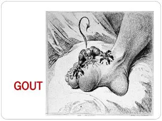
CLINICAL MANIFESTASIONS Gout has two clinical phases. The first phase is characterized by intermittent acute attacks that spontaneously resolve, typically over a period of 7 to 10 days, with asymptomatic periods between attacks. With inadequately treated hyperuricemia, transition to the second phase can occur, manifested as chronic tophaceous gout, which often involves polyarticular attacks, symptoms between attacks, and crystal deposition (tophi) in soft tissues or joints. Although the prevalence of tophaceous gout varies among populations, in one study, tophi were detected in three quarters of patients who had had untreated gout for 20 years or more. (Gutman AB. The past four decades of progress in the knowledge of gout, with an assessment of the present status. Arthritis Rheum 1973;16:431-45.) Acute arthritis is the most frequent early clinical manifestation of gout. Usually, only one joint is affected initially, but polyarticular acute gout can occur in subsequent episodes. The metatarsophalangeal joint of the first toe is often involved, but tarsal joint, ankles and knees are also commonly affected. Especially in elder patients or in advanced disease, finger joints may be involved. (Harrison’s Principles of Internal Medicine, 17 Edition Volume 2, p.2165) Gout occurs most commonly in the big toe because uric acid is sensitive to temperature changes. At cooler temperatures, uric acid turns into crystals. Since the toe is the part of the body that is farthest from the heart, it’s also the coolest part of the body – and, thus, the most likely target of gout. However, gout can affect any joint in the body. (http://www.foothealthfacts.org/footankleinfo/gout.htm)
The first episode of acute gouty arthritis frequently begins at night with dramatic joint pain and swelling. Joints rapidly become warm, red, and tender, with a clinical appearance that often mimics cellulitis. Early attacks tend to subside spontaneously within 3-10 days, ad most patients have interval of varying length with no residual symptoms until next episode. Several events may precpitate acute gouty arthritis: dietary excess, trauma, surgery, excessive ethanol ingestion, hypouricemic therapy, and serious medical illness such as myocardial infarction and stroke. (Harrison’s Principles of Internal Medicine, 17 edition Volume 2, p.2165) Stadium artritis gout menahun (chronic gout arthritis) umumnyaterjadipadapasien yang mengobatidirisendiri. Biasanyadisertai tophus danpoliartikular. Tophus seringpecahdansulitsembuhdenganobat, bisaterjadiinfeksisekunder. Lokasi tophus yang paling seringadadi MTP-1, cupingtelinga, olekranon, tendon achillesdanjaritangan. Pada stadium inikadang-kadangdisertaibatusalurankemihdanpenyakitginjalmenahun. (BukuIlmuPenyakitDalam FK UI, Edisi 5 Jilid 3, p.2559)
PATOPHYSIOLOGY Gout is a type of inflammatory arthritis, occurs when final metabolite of purine, uric acid, crystallizes in the form of monosodium urate in synovial fluid. These crystals then trigger a local immune-mediated inflammatory reaction, with one of the key proteins in the inflammatory cascade being interleukin 1β. (Richette P, Bardin T (January 2010). "Gout". Lancet 375 (9711): 318–28. ) Kristal uratdapatmengaktifkansistemkomplemenmelaluijalurklasikdanjaluralternatif. Melaluijalurklasik, terjadiaktivasikomplemen C1 tanpaperanimunoglobulin. Padakadar MSU meninggi, aktivasisistemkomplemenmelaluijaluralternatifterjadiapabilajalurklasikterhambat. Aktivasikomplemenmenyebabkanpeningkatanaktivitasproseskemotaksisselneutrofil, vasodilatasisertapengeluaransitokin IL-1 dan TNF, yang menyebabkanperadangan. Tujuanperadanganpada gout adalahsebagaireaksipertahanantubuh non-spesifik, untukmenetralisiragenpenyebab, danmencegahperluasanagenpenyebab. (BukuIlmuPenyakitDalam FK UI, Edisi 5 Jili 3, p.2557) Penurunanasamurat serum dapatmencetuskanpelepasankristal monosodium uratdaridepositnyadisinoviumatautofi. Pelepasankristal MSU akanmerangsangprosesinflamasidenganmengaktifkankomplemenmelaluijalurklasikmaupunalternatif. (HidayatR.Goutdanhiperurisemia. Medicinus 2009;Vol.22.)
Gout occurs most commonly in the big toe because uric acid is sensitive to temperature changes. At cooler temperatures, uric acid turns into crystals. Since the toe is the part of the body that is farthest from the heart, it’s also the coolest part of the body – and, thus, the most likely target of gout. However, gout can affect any joint in the body. (http://www.foothealthfacts.org/footankleinfo/gout.htm) See gout patophysiology.
DIAGNOSIS The diagnostic standard remains synovial fluid or tophus aspiration with identification of negatively birefringent monosodium urate crystals under polarizing microscopy. Crystals are detectable during attacks and also potentially between attacks, primarily in previously inflamed joints in patients with hyperuricemia. (Zhang W, Doherty M, Pascual E, et al. EULAR evidence based recommendations for gout. Part I: Diagnosis. Report of a task force of the EULAR Standing Committee for International Clinical Studies Including Therapeutics (ESCISIT). Ann Rheum Dis 2006;65:1301-11.) Even if the clinical appearance strongly suggests gout, the diagnosis should be confirmed by needle aspiration of acutely or chronically involved joints or tophaceous deposits. During acute gouty attacks, strongly birefringent needle-shaped MSU crystals with negative elongation are typically seen both intracellularly and extracellularly. Synovial fluid count are elevated from 2000 to 60.000/μL. Bacterial infection can coexist with urate crystals synovial fluid; if there is any suspicion of septic arthritis, joint fluid must also be cultured. (Harrison’s Principles of Internal Medicine, 17 Edition Volume 2, p.2166) See gout crystal patology. However, crystal evaluation is not performed routinely in clinical practice.
Hyperuricemia may not be present during acute gout attacks and therefore may not be a helpful criterion for diagnosis. A typical presentation that is strongly suggestive of the diagnosis includes rapid development of severe pain (i.e., within 24 hours), erythema, and swelling in a characteristic joint distribution — for example, in the first metatarsophalangeal joint (podagra). In a population with a 0.5% prevalence of gout overall, a patient with hyperuricemia and this presentation has an 82% chance of having gout. (Zhang W, Doherty M, Pascual E, et al. EULAR evidence based recommendations for gout. Part I: Diagnosis. Report of a task force of the EULAR Standing Committee for International Clinical Studies Including Therapeutics (ESCISIT). Ann Rheum Dis 2006;65:1301-11.) Kombinasidaripenemuan-penemuandibawahinidapatdipakaiuntukmenegakkan diagnosis: Riwayatinflamasiklasik arthritis monoartikulerpadasendi MTP-1 Diikuti stadium asimptomatic Resolusisinovitis yang cepatdenganpengobatankolsikin Hiperurisemia Kadar asamurat normal tidakdapatmenghindari diagnosis gout. Logan dkkmendapatkan 40% pasien gout dengankadarasamurat normal. Hasilpenelitiandidapatkansebanyak 21% arthritis gout denganasamurat normal. Kriteriauntukpenyembuhanakibatpengobatandengankolsikinadalahhilangnyagejalaobjektifinflamasipadasetiapsendidalamwaktu 7 hari. (BukuIlmuPenyakitDalam FK UI, Edisi 5 Jilid 3, p.2559)
The differential diagnosis of acute gout includes other crystal-induced arthritides (e.g., calcium pyrophosphate dihydrate) and a septic joint. Joint aspiration with Gram’s staining and culture must be performed if a septic joint is suspected, even if monosodium urate crystals are identified. Older adults, particularly women, may present with polyarticular involvement, which may be mistaken for rheumatoid arthritis; a tophus may be mistaken for a rheumatoid nodule. Tophaceous deposits that are not clinically apparent may be visualized by plain radiography or another imaging method. A diagnosis of gout should prompt evaluation for potentially modifiable risk factors (e.g., dietary habits) and associated coexisting illnesses (e.g., hypertension and hyperlipidemia) that may require intervention.
PHARMACOLOGIC TREATMENT The mainstay of treatment during acute attack is the administration of anti-inflammatory drugs such as NSAIDs, colchicine, or glucocorticoids. NSAIDs are most often used in individuals without complicating comorbid conditions.. Both colchicine and NSAIDs may be poorly tolerated and dangerous in the elderly and in the presence of renal insufficiency and gastrointestinal disorders. In attack involving one or two joints, intraarticularglucocorticoid injections may be preferable and effective. Ice pack applications and rest of the involved joints can be helpful. (Harrison’s Principles of Internal Medicine, 17 Edition Volume 2, p.2166) The choice of agent, dose, and duration of therapy is guided by consideration of coexisting illnesses that preclude the safe use of a particular regimen, as well as the severity of the gout. Adjunctive measures include applying ice to and resting the affected joint. (Schlesinger N, Detry MA, Holland BK, et al. Local ice therapy during bouts of acute gouty arthritis. J Rheumatol 2002; 29:331-4.) NSAIDs and colchicine are first-line agents for acute attacks. (Zhang W, Doherty M, Bardin T, et al. EULAR evidence based recommendations for gout. Part II: Management. Report of a task force of the EULAR Standing Committee for International Clinical Studies Including Therapeutics (ESCISIT). Ann Rheum Dis 2006;65:1312-24.)
NSAIDs given in full anti-inflammatory doses are effective in ~90% of patients, and the resolution of signs and symptoms usually occurs in 5-8 days. The most effective drugs are any of those with a short half-life and include indomethacin, 25-50 mg tid; ibuprofen, 800 mg tid; or diclofenac 50 mg tid. (Harrison’s Principles of Internal Medicine, 17 Edition Volume 2, p.2166) In patients with increased risk of peptic ulcers, bleeds or perforations, co-prescription of gastro-protective agents should follow standard guidelines for the use of NSAIDs. (Kelsey M. Jordan, J. Stewart Cameron, et al. British Society for Rheumatology and British Health Professionals in Rheumatology Guideline for the Management of Gout. Oxford University 2007.) Colchicine given orally is a traditional and effective treatment, if used early in the attack. One to two 0,6 mg tablets can be given every 6-8 h over several days with subsequent tapering. This is generally better tolerated than the formerly advised regimen. The drug must be stopped promptly at the first sign of loose stools, and symptomatic treatment must be given for the diarrhea. Intravenous colchicine is occasionally used, e.g., as pre- or postoperative prophylaxis in 1 – to 2-mg doses when patients cannot take medications orally. Life threatening colchicine toxicity and sudden death have been described with the administration of >4mg/d IV. The IV colchicine should be given slowly through an established venous line over 10 min in a soluset. The total doses should never exceed 4 mg. (Harrison’s Principles of Internal Medicine, 17 Edition Volume 2, p.2166)
Oral colchicine has long been used, although it has only recently (in 2009) been approved by the Food and Drug Administration (FDA) for use in patients with acute gout. In a randomized trial, colchicine (at a dose of 1.2 mg at the onset of a flare, followed by 0.6 mg 1 hour later) was significantly more likely than placebo to result in a reduction in pain of 50% or more 24 hours later (rates, 37.8% and 15.5%, respectively). This regimen had efficacy similar to that of a high-dose regimen (1.2 mg, then 0.6 mg per hour for 6 hours), with fewer gastrointestinal side effects. This study did not address treatment after the first 24 hours. (Terkeltaub RA, Furst DE, Bennett K, Kook KA, Crockett RS, Davis MW. High versus low dosing of oral colchicine for early acute gout flare: twenty-four-hour outcome of the first multicenter, randomized, double-blind, placebo-controlled, parallelgroup, dose-comparison colchicine study. Arthritis Rheum 2010;62:1060-8.) Oral glucocorticoids such as prednisone, 30-50 mg/d as the initial dose and gradually tapered with the resolution of the attack can be effective in polyarticular gout. For single of few involved joints intraarticulartriamcinoloneacetonide, 20-40 mg, or methylprednisolone, 25-50 mg, have been effective and well tolerated. ACTH as intramuscular injection of 40-80 IU in a single dose or every 12 h for 1-2 days can be effective in patients with acute polyarticular refractory gout or in those with a contraindication for using colchicine or NSAIDs. (Harrison’s Principles of Internal Medicine, 17 Edition Volume 2, p.2166)
When the use of NSAIDs or colchicine is poorly tolerated or contraindicated, glucocorticoids or corticotropin may be used, although evidence for the use of intraarticular and intramuscular glucocorticoids and corticotropin is limited by a lack of data from blinded, randomized, placebocontrolled trials. (Zhang W, Doherty M, Bardin T, et al. EULAR evidence based recommendations for gout. Part II: Management. Report of a task force of the EULAR Standing Committee for International Clinical Studies Including Therapeutics (ESCISIT). Ann Rheum Dis 2006;65:1312-24.) Kortikosteroiddan ACTH diberikanapabilakolsikindan OAINS tidakefektifataumerupakankontraindikasi. Pemakaiankortikosteroidpada gout diberikan oral atauparenteral. Indikasipemberianadalahpadaartritis gout polyartikular. (BukuIlmuPenyakitDalam FK UI, Edisi 5 Jilid 3, p.2559-2560) See pharmacologic table.
Ultimate control of gout requires correction of the basic underlying defect, the hyperuricemia. Attempts to normalize serum uric acid to <300-360 μmol/L (5.0-6.0 mg/dL) to prevent recurrent gouty attacks and eliminate tophaceus deposits entail a commitment to long-term hypouricemic regimens and medications that generally are required for life. Hypouricemic therapy should be considered when, as in most patients, the hyperuricemia cannot be corrected by simple means (control of body weight, low-purine diet, increase in liquid intake, limitation of ethanol use, and avoidance of diuretics). The decision to initiate hypouricemic therapy is usually made taking into consideration the number of acute attacks (urate lowering therapy may be cost effective after two attacks), serum uric acid levels (progession is more rapid in patients with serum uric acid >535 μmol/L [9.0 mg/dL]), patient’s willingness to commit to lifelong therapy, or presence of uric acid stones. Urate-lowering therapy should be initiated in any patient who already has tophi or chronic goutly arthritis. (Harrison’s Principles of Internal Medicine, 17 Edition Volume 2, p.2166) The purpose of lowering serum urate levels is to prevent acute flares and development of tophi. However, gout does not develop in all patients with hyperuricemia, and antihyperuricemic therapies are not without risk. Recommendations that are based on both consensus and evidence support the consideration of urate-lowering therapy in patients with hyperuricemia who have at least two gout attacks per year or tophi (as determined by either clinical or radiographic methods). (Mikuls TR, MacLean CH, Olivieri J, et al. Quality of care indicators for gout management. Arthritis Rheum 2004;50:937-43.)
However, the severity and frequency of flares, the presence of coexisting illnesses (including nephrolithiasis), and patient preference are additional considerations. (Zhang W, Doherty M, Bardin T, et al. EULAR evidence based recommendations for gout. Part II: Management. Report of a task force of the EULAR Standing Committee for International Clinical (Studies Including Therapeutics (ESCISIT). Ann Rheum Dis 2006;65:1312-24.) Urate-lowering therapy should not be initiated during acute attacks but rather started 2 to 4 weeks after flare resolution, with a low initial dose that is increased as needed over a period of weeks to months, and with close monitoring of urate levels, renal function, and adverse effects. The dose should be adjusted as necessary to maintain a serum urate level below 6 mg per deciliter (357 μmol per liter), which is associated with a reduced risk of recurrent attacks and tophi. (Perez-Ruiz F, Lioté F. Lowering serum uric acid levels: what is the optimal target for improving clinical outcomes in gout? Arthritis Rheum 2007;57:1324-8.)
Kontrolhiperurisemiadilakukandengan diet rendahpurin, sertamenghindariobat-obatan yang meningkatkankadarasamuratseruterutamadiuretik. Selanjutnyadiperlukanurate lowering agent sepertigolonganxanthineoxidase inhibitor, maupunuricosuric agent, dengancatatantidakbolehdimulaipadasaatseranganakut. Padahiperurisemiaasimptomatikterapifarmakologikdimulaijikakadarasamurat serum >9mg/dL. Sedangkanpadapenderita gout telahdiketahuibahwapemberianurate lowering agent jugamenjadifaktorpencetusseranganakut, sehinggadiberikanjugakolsikindosisprevensi 0,6 mg 1-3 kali perhari, atau OAINS dosisrendah, dandimulaisetelahtidakadanyatanda-tandainflamasiakut. (HidayatR.Goutdanhiperurisemia. Medicinus 2009;Vol.22.) Three classes of drugs are approved for lowering urate levels: xanthineoxidase inhibitors, uricosuric agents, and uricase agents. (Becker MA, Schumacher HR, Espinoza LR, et al. The urate-lowering efficacy and safety of febuxostat in the treatment of the hyperuricemia of gout: the CONFIRMS trial. Arthritis Res Ther 2010;12:R63.) Although allopurinol has an acceptable side-effect profile in most patients, a mild rash develops in approximately 2%. The majority of patients receive 300 mg of allopurinol daily, but this dose is often inadequate to achieve target urate levels. Daily doses up to 800 mg may be used in patients with normal renal function. The dose is typically reduced in patients with renal impairment, owing to concerns about an increased risk of hypersensitivity in such patients. (Dalbeth N, Kumar S, Stamp L, Gow P. Dose adjustment of allopurinol according to creatinine clearance does not provide adequate control of hyperuricemia in patients with gout. J Rheumatol 2006;33: 1646-50.)
Jenisurate lowering agent yang pertamayaitugolonganxanthineoxidase inhibitor dengancarakerjapenghambatanoksidasihipoxantinmenjadixantin, danxantinmenjadi uric acid. Sedangkanjenisurate lowering agent yang keduayaitugolonganuricosuric agent, bekerjadengancaramenghambatreabsorpsiuratditubulusrenalis. (HidayatR.Goutdanhiperurisemia. Medicinus 2009;Vol.22.) Allopurinolsebagaixanthineoxidase inhibitor diberikanmulaidosis 100 mg/haridandinaikkantiapminggusampaitercapai target (rata-rata diperlukan minimal 300 mg/hari). Padagangguanfungsiginjaldosisharusdisesuaikan. (HidayatR.Goutdanhiperurisemia. Medicinus 2009;Vol.22.) Golonganuricosuric agent, probenesiddengandosis 0,5-3 gram dibagi 2-3 kali perhari. Sedankansulfinpirazondiberikandengandosis 300-400 mg dibagi 3-4 kali perhari. Pemakaianobaturicosuricinilebihdiindikasikanpadakeadaandenganekskresiasamuratdiurin <800 mg perhari, dandenganfungsiginjalmasihbaik (creatinin clearance >80 mL/menit). (HidayatR.Goutdanhiperurisemia. Medicinus 2009;Vol.22.) Urate-lowering drugs are generally not initiated during acute attacks but after the patient is stable and low-dose colchicine has been initiated to decrease the risk of flares that often occur with urate lowering. Colchicine prophylaxis in doses of 0,6 mg one to two times daily is usually continued, along with the hypouricemic therapy, until the patient is normouricemic and without gouty attacks for 6 months or as long as tophi are present. (Harrison’s Principles of Internal Medicine, 17 Edition Volume 2, p.2166)
See pharmacologic table 2. Urate-lowering therapy, which reduces the risk of gout attacks in the long term, can trigger attacks in the early period after its initiation, presumably as a result of mobilization of bodily urate stores. (Borstad GC, Bryant LR, Abel MP, Scroggie DA, Harris MD, Alloway JA. Colchicine for prophylaxis of acute flares when initiating allopurinol for chronic gouty arthritis. J Rheumatol 2004;31: 2429-32.) (Becker MA, Schumacher HR Jr, Wortmann RL, et al. Febuxostat compared with allopurinol in patients with hyperuricemia and gout. N Engl J Med 2005;353: 2450-61.) Penurunanasamurat serum dapatmencetuskanpelepasankristal monosodium uratdaridepositnyadisinoviumatautofi. Pelepasankristal MSU akanmerangsangprosesinflamasidenganmengaktifkankomplemenmelaluijalurklasikmaupunalternatif. (HidayatR.Goutdanhiperurisemia. Medicinus 2009;Vol.22.)
NON-PHARMACOLOGIC TREATMENT Observational data indicate that nonpharmacologic approaches, such as avoiding alcohol or modifying one’s diet, can reduce serum urate levels but may not be sufficient to control established gout. (Zhang W, Doherty M, Bardin T, et al. EULAR evidence based recommendations for gout. Part II: Management. Report of a task force of the EULAR Standing Committee for International Clinical Studies Including Therapeutics (ESCISIT). Ann Rheum Dis 2006;65:1312-24.) Low purine diet. Food never contains uric acid. Some food contains purines, which are a source of uric acid. See food containing purines table. Alcohol promotes hyperuricemia because of increased urate production and decreased uric acid excretion. Excessive alcohol consumption accelerates hepatic breakdown of ATP to increase urate production. Alcohol consumption induce hyperlacticacidemia, which blocks uric acid secretion. (Harrison’s Principles of Internal Medicine, 17 Edition Volume 2, p.2446) In one randomized trial involving persons without gout, 500 mg of vitamin C per day for 2 months resulted in serum urate levels that were 0.5 mg per deciliter (30 μmol per liter) lower than in those receiving placebo. (Huang HY, Appel LJ, Choi MJ, et al. The effects of vitamin C supplementation on serum concentrations of uric acid: results of a randomized controlled trial. Arthritis Rheum 2005;52:1843-7.)
The intake of dairy milk reduced serum urate levels by approximately 10% during a 3-hour period in a small, randomized, crossover trial involving healthy volunteers. (Dalbeth N, Wong S, Gamble GD, et al. Acute effect of milk on serum urate concentrations: a randomised controlled crossover trial. Ann Rheum Dis 2010;69:1677-82.) Losartan and fenofibrate, which have uricosuric effects, may be considered in patients with gout who have hypertension or hypertriglyceridemia. (Takahashi S, Moriwaki Y, Yamamoto T, Tsutsumi Z, Ka T, Fukuchi M. Effects of combination treatment using anti-hyperuricaemic agents with fenofibrate and/or losartan on uric acid metabolism. Ann Rheum Dis 2003;62:572-5.)