Download
1 / 24
470 likes | 1.66k Views
COMPARATIVE VERTEBRATE ANATOMY LECTURE. BSPT 22 BSPT 23 INSTRUCTOR : I.A.RONIO. TYPES OF CELL DIVISION MITOSIS-duplication division; occurs in ___. MEIOSIS-reduction division; occurs in ____.

E N D
COMPARATIVE VERTEBRATE ANATOMY LECTURE BSPT 22 BSPT 23 INSTRUCTOR : I.A.RONIO
TYPES OF CELL DIVISION MITOSIS-duplication division; occurs in ___. MEIOSIS-reduction division; occurs in ____. subdivisions: Meiosis I -simple mitotic division that produces two identical cells, each with a complete set of chromosomes. Meiosis II -the two new identical cells produced in the first stage undergo reduction and division, producing two new cells with half of the complete set of chromosomes of the original parent cell.
GENERAL FEATURES OF CHORDATE DEVELOPMENT A. The Chordate Egg According to amount of yolk: 1. ISOLECITHAL 2. TELOLECITHAL with TOTAL UNEQUAL CLEAVAGE 3. TELOLECITHAL with MEROBLASTIC CLEAVAGE B. The Cleavage of the Egg and the Formation of Blastula 1. HOLOBLASTIC EQUAL CLEAVAGE 2. HOLOBLASTIC UNEQUAL CLEAVAGE 3. MEROBLASTIC CLEAVAGE C. Formation of the Gastrula 1. In Amphioxus Type Eggs 2. In Amphibian Type Eggs 3. In Meroblastic Eggs
D. Formation of the 3rd Germ Layer, the Neural Tube, and the Notochord 1. In Amphioxus 2. In Vertabrates E. Further History of the Mesoderm F. The Fate of the Ectoderm G. The Ftae of the Endoderm H. The Fate of the Mesoderm and the Formation of the Mesenchyme 1. MESENCHYME 2. The fate of the EPIMERE 3. The Fate of the MESOMERE 4. The Fate of the HYPOMERE 5. The Products of the Mesenchyme I. The Homology of the Germ Layers
Steps in Early Craniate Development • Sperm cell and egg cell nuclei fusion (process is known as__________ that forms the zygote or the________) ->morula (process is known as___________) ->blastula (process is known as__________) ->gastrula (process is known as__________) ->neurula (process is known as__________) ->embryo (that will undergo organogenesis to form _________).
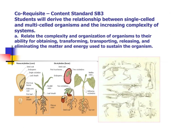
Ovum Types as to Amount of Yolk • Microlecithal (amphioxus;placental mammals) • Mesolecithhal (amphibians;lampreys;ganoid fishes) • Macrolecithal (birds;sharks;reptiles;teleosts)
Ovum Types As To Yolk Distribution • Isolecithal Egg present in amphioxus and mammals. • Telolecithal Egg with total cleavage present in amphibians and some fishes. • Telolecithal Eggs with meroblastic cleavage present in some fishes, reptiles, birds, and in some mammalian eggs.
Cleavage Patterns • Holoblastic Equal Cleavage happens among isolecithal eggs. • Holoblastic Unequal Cleavage happens in telolecithal eggs having total cleavage. • Meroblastic Cleavage happens in telolecithal eggs wherein the only the germinal disk undergoes cleavage.
EARLY CLEAVAGE IN AMPHIOXUS During the earliest cleavages, growth does not occur. In fact, as nutritive materials are used to power the earliest processes, the embryo may actually decrease in size. Cleavage planes pass entirely through the egg. They are holoblastic. The first two are meridional, giving the four-cell stage. The third is equatorial. The third cleavage is not exactly perfectly distributed. Lower, yolkier cells are somewhat larger.
GASTRULATION Due to faster growth of cells at the position of the prospective blastopore, the surface of the region increases. This causes a dimpling in, or INVAGINATION. In addition to invagination, we also have INVOLUTION, a movement of cells inward s fast as they are produced. This multiplication and involution takes place most rapidly at the dorsal lip of the blastopore. The dorsal lip of the blastopore is an important organizing region for the embryo. In the following two-dimensional drawing, realize that the blastopore is representing a circular opening.
Internal View of the Primitive Streak During gastrulation in birds and mammals, epiblast cells converge at the midline and ingress at the primitive streak. Ingression of these cells results in formation of the mesoderm and replacement of some of the hypoblast cells to produce the definitive endoderm.
Neurulation • In vertebrates results in the formation of the neural tube, which gives rise to both the spinal cord and the brain. • Neural crest cells are also created during neurulation. • Neural crest cells migrate away from the neural tube and give rise to a variety of cell types, including pigment cells and neurons. • Neurulation begins with the formation of a neural plate, a thickening of the ectoderm caused when cuboidal epithelial cells become columnar. • Changes in cell shape and cell adhesion cause the edges of the plate fold and rise, meeting in the midline to form a tube. • The cells at the tips of the neural folds come to lie between the neural tube and the overlying epidermis. • These cells become the neural crest cells. • Both epidermis and neural plate are capable of giving rise to neural crest cells. • The notochord is necessary in order to induce neural plate formation.
Neurulation in Amphioxus After gastrulation, we enter the stages of neurulation, where a number of processes occur simultaneously. It is impossible to follow all of them at once, so we will try to break them down. First in transverse section we will follow the process of formation of the neural tube and somites. As endoderm thickens, it breaks away from the epidermal ectoderm and comes to sink in as the NEURAL PLATE. (This is different from what we will see in vertebrates where the neural ectoderm rolls up on itself.) Neural plate formation is induced by the notochord tissue.
Neurulation in Mammals and Birds The region where neural tube closure begins varies between different classes of vertebrates. In amphibians, the neural tube closes almost simultaneously along its entire length. In birds, the neural tube closes in the anterior to posterior direction, as Hensen's node regresses. In mammalians, neurulation is similar to that of birds, however the bulky anterior neural plate seems to resist closure - the middle of the tube closes first, followed by both ends.
Somites during Neurulation During neurulation, somites form in pairs flanking the neural tube. Somites are blocks of cells that form a segmental pattern in the vertebrate embryo. Somites produce cells that become vertebrae, ribs, muscles, and skin.
To Be Continued... THANK YOU