Download
1 / 42
510 likes | 901 Views
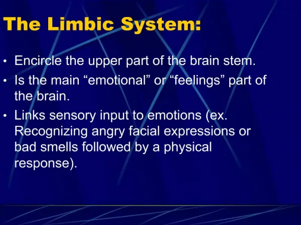
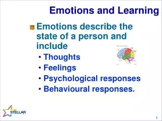
The Limbic System and Cerebral Circuits for Emotions, Learning and Memory. Introduction to the Limbic System. Anatomically refers to areas surrounding the diencephalon (limbus = border) and bordering the cerebral cortex.

E N D
The Limbic System and Cerebral Circuits for Emotions, Learning and Memory
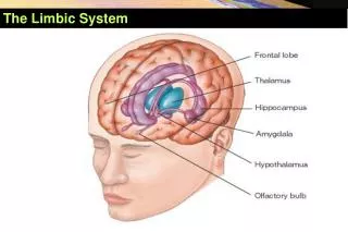
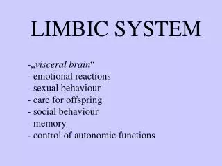
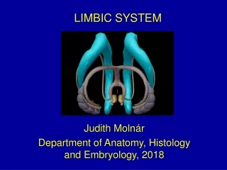
Introduction to the Limbic System • Anatomically refers to areas surrounding the diencephalon (limbus = border) and bordering the cerebral cortex. • The “C”-shaped hippocampal formation and includes the amygdala, cingulate and parahippocampal cortices. The key to learning, memory, and behavior (including emotional behavior) – of paramount importance in psychiatry.
Anatomical Location + discussion of components. • Structure and function of the hippocampal formation. • The Amygdaloid nucleus complex and the function of its 3 components. • Links between the limbic system and effector (behaviour) systems. • Links between neurotransmitter-specific projection systems and the limbic system. • Regional Anatomy of these structures.
Anterior section: Amygdala(Fig. 16-13) Divisions or nuclei of amygdala
Posterior section: Hippocampus, Fornix(Fig. 16-15) Divisions or nuclei of hippocampal formation
Midsagital section(Fig. 16-19) Note the mammillary body, mammillothalamic tract, and fornix (body).
Parasagital section(Fig. 16-19) Note the hippocampal formation, fornix, and amygdala
Anatomical Location and Overview. • Limbic association cortex – surrounding diencephalon -medial + inferior (orbital) surface cingulate gyrus, parahippocampal gyrus, orbital gyrus, temporal pole.
Figs. 16-2 and 3 Limbic System: Cortical Areas Note: surrounding diencephalon, medial + inferior (orbital) surface [cinglulate gyrus, parahipp gyrus, orbital gyrus, temporal pole]. These cortices are near other association cortices and project to:
B. the Hippocampal Formation and Amygdala (Fig. 16-1). Note the “C” shape, along with the major output paths for the hippocampus: the fornix. Note the mammillary body: it is part of what structure? Refer to Table 16-1 – Lists all the components of the limbic system.
II. Hippocampal Formation: Components A subcortical structure composed of allocortex. A central function: Consolidation of STMs into LTMs (+ many other limbic functions through complex interconnections). Note the 3 components
Neocortex and Allocortex(Fig. 16-12) More on these 3 layers later
Hippocampal Formation: • Circuitry (Fig. 16-4). • Components and structure – • a banana-shaped structure with its • components (dentate, hipp, • subiculum) folded upon one • another like a “jelly roll”. • Inputs are from entorhinal • cortex, which collects info • from other association areas • dentate gyrus • hipp formation + subculum • output to fornix and • also back to entorhinal cortex • (See Fig. 16-6).
Hippocampal Formation: Input and Output (Fig. 16-6) Afferent Efferent
Serial and Parallel Processing of Hippocampal Circuits (Fig. 16-5)
Afferents Efferents Hippocampal Circuits Contralateral Hippocampus 3. 4. (septal-hippocampal pathway) Affects - Theta rhythm (4-8Hz) (Hippocampal commissure) Cortex F F o o r r n n i i x x branch (Cortico-entorhinal projections) HIPPOCAMPUS (Precommissural) (Postcommisural Septal nuclei 1. & 2. (Perforant & alvear path) Branch) Mammillary Body ERC/Sub (PHG)
The Hippocampus Dentate Complex(HC-DG)Afferent Pathways 3. (septo-hippocampal path - thru fornix) Septal nuclei Pyramidal cell (CA1,2) (schaffer collaterals) (Also note: this efferent path closes the HC circuit loop!) Pyramidal cell (CA3) 2. (alvear path) Dentate gyrus (granule cells) PHG (ERC, Sub) (mossy fibers) 1. (perforant path) Perforant Pathway: PHG (ERC) --> DG Also …. Alvear Pathway: PHG --> CA1 Septo-hippocampal path (via fornix): Septal nuclei --> DG Hippocampal commissure (connects bilateral hippocampi)
Papez’ Circuit: Fornix mammillary bodies mammillothalamic tract Hippocampal formation Thalamic nuclei Entorhinal cortex Cingulate gyrus Input for memory consolidation
Korsokoff’s Syndrome: thiamine deficiency (i.e., from alcoholism) degeneration of mammillary bodies. • Other output: via entorhinal cortex to a number of association areas, involve the prefrontal cortex (control of mood and behaviours).
C. Anatomy and Information Flow in Greater Detail. Cytoarchitechture dimensions of hippocampus (Ammon’s horn): CA1, CA2, CA3, CA4 (hilus) [CA = coronus ammonis].
Medial Temporal Circuitry –See Fig. 16-18 for Corresponding Anatomy Section Pyramidal cells Sub CA 1 PRC Pyramidal cells Schaffer collaterals ERC Granule cells Pyramidal cells PHC DG CA 3 Fornix Mossy fibers Hippocampus (HC) proper : Dentate Gyrus (DG), CA3, CA1, and Subiculum (Sub) Adjacent MTL cortices : Entorhinal (ERC), Perirhinal (PRC) Parahippocampal (PHC)
The HippocampusCA fields A) Lateral ventricle B) Ependymal glia C) Alvear layer 1. Polymorph Layer (pyramidal axon) (pyramidal cell body) 2. Pyramidal Layer (pyramidal dendrite) 3. Molecular Layer A) Lateral Ventricle, B) ependymal glia (ventricular surface), C) Alvear Layer, (pyramidal axons) 3 layers of hippocampus (archicortex): Polymorph Layer (pyramidal axons & basket cells (-)) Hippocampal pyramidal layer (pyramidal cell bodies) Molecular Layer (pyramidal dendrites)
Taxonomy of Long-term Memory Systems Squire L, Zola S PNAS 1996;93:13515-13522 Adapted from Squire, Knowlton 1994
Patient H.M. and the Human MTL • Suffered head injury @age 9 • Developed severe epilepsy • Surgeon surgically removed the medial temporal lobe bilaterally • HM suffered severe anterograde and temporally graded retrograde amnesia • Spared skill learning (Corkin, Amaral, Gonzalez, Johnson and Hyman J. Neuro, 1997) (Scoville and Milner, 1957)
III. Amygdala • Almond-shaped • Major function: Responding to stimuli with an emotional component. • 3 Nuclear components: A. Basolateral – attaching emotional significance to a stimulus. Sensory cortices (higher-order) BL limbic associaation cortex, prefrontal cortex, hippocampal formation (for learning emotional significance).
B. Central Nucleus – mediates emotional responses. Visuaosensory input from solitary, parabrachial nuclei CN dorsal motor n. of X, other parasympathetic n. reticular formation and hypothalamus autonomic responses. C. Corticomedial nuclei – mediates behaviors triggered by olfactory stimuli. Olfactory bulb CM hypothalamus (lateral zone) regulation of “appetitive behaviours”; i.e., eating in response to smells.
IV. Links Between Limbic System and Effector (Behavioural) Systems • Neuroendocrine – by amygdala (central and CM) via paraventricular n. of hypothalamus. • Autonomic – by amygdala (central n.?) lateral hypothalamus descending pathways via autonomic nuclei. • Somatic Motor – by several limbic components reticular formation stereotypic behaviour via reticulospinal tract.
V. Links Between Neurotransmitter-Specific Projection Systems and Limbic Systems This projection appears to be vital for normal thoughts, moods, and behaviours. We have known this empirically because of known behavioural disturbances in animals with lesions of these systems and neurotransmitter-activating or –blocking drugs seem to help humans manage behavioural aberrations to some degree.
Dopamine: Excessive transmission in limbic structures may contribute to schizophrenia. 5-HT: Ascending projections control mood; increase appears to help in many forms of anxiety and depression (mood disorders). Norepinephrine: Excessive transmission may contribute to anxiety and aggression. Under-reaction, along with that of 5-HT, may contribute to depression. Acetylcholine: Important in cognition, activating a number of neocortical, as well as limbic areas – the 1st system to degenerate in Alzheimer’s Disease.
DA NE Fig. 2-2 5-HT
VI. Regional Anatomy See outline and stained sections of structures (Figs. 16-13, 15, 19).