Download
1 / 35
350 likes | 466 Views
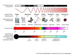
X-rays are electromagnetic waves (like light) and have a wavelength about the size of atoms. From https://askabiologist.asu.edu/quick-class-x-ray-crystallography. X-rays.

E N D
X-rays are electromagnetic waves (like light) and have a wavelength about the size of atoms. From https://askabiologist.asu.edu/quick-class-x-ray-crystallography
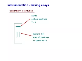
X-rays • X rays are electromagnetic radiation ranging in wavelength from about 100 A to 0.01 Å (1 Å is equivalent to about 10-8 cm (about 4 billionths of an inch). • The shorter the wavelength of the X ray, the greater is its energy and its penetrating power. • A mixture of many different wavelengths is known as “white” X rays, as opposed to “monochromatic” X rays, which represent only a single wavelength. Wilhelm Conrad Röntgen, 1845-1923 The hand of Albert von Kolliker, 1817-1905. Introduced many microscopic mechniques to advance Histology.
Rotating anode generator Rotating anode
Simplified phase diagram for crystallization Different techniques traverse the crystallization space differently.
0.9 Å 1.4 Å Beam stop shadow Diffracts to beyond 0.85 Å. In this image ~5000 data points alone are visible. The total data set at this resolution has over 1 million data points.
2Fo-Fc 1.0s Fo-Fc 4s Fo-Fc 2s Fo-Fc 5s Fo-Fc 3s
For more signal, need more intensity The first synchrotron discovered, the Crab Nebula (about 6500 light years away)
A synchrotron accelerates and stores particles (electrons or protons) moving at speeds close to that of light. As the particles loose energy they give of electromagnetic radiation. The particles are steered by magnetic fields. Electromagnetic radiation (photons) is not affected by these fields and is emitted at the tangent to the change in direction. Insertion devices (undulators and wigglers) ‘amplify’ this radiation
Synchrotron radiation is 109 times More brilliant than the sun and about 100 million miles closer
Linear accelerator Booster Ring Synchrotron Beamline
Where? NSLS APS CHESS SNS SSRL ILL JAERI Synchrotron X-ray sources we are actively using Neutron sources we are actively using
More candles provide more light “Evolutionary technology” Massimo Catarinella
A light bulb also provides light. But it is an example of revolutionary technology It changed the way we do things
We are here now! Synchrotron radiation is 109 times More brilliant than the sun and about 100 million miles closer
The Linear Coherent Light Source XFEL X-ray Free Electron Lasers (XFEL’s) are revolutionary sources of X-ray radiation They are changing the way and the kind of X-ray based science we can do Photo courtesy of SLAC National Accelerator Laboratory
Moving electrons can emit X-rays through several processes, in our case the emission (synchrotron radiation) is caused by changing direction due to a magnetic field The force felt by a charged particle (an electron in this case) in a magnetic field is perpendicular to the direction of the field and to the direction of the particle's velocity. The net effect of this is to cause the particle to spiral around the direction of the field. Since circular motion represents acceleration (i.e., a change in velocity), electrons radiate photons (which include X-rays) of a characteristic energy, corresponding to the radius of the circle Image from NASA
The first part of an XFEL, the injector, is used to generate electrons In the LCLS case (an XFEL at Stanford) the injector cathode is a highly polished copper plate. A short burst of light from a drive laser hits the cathode's shiny surface and produces one electron for every 100,000 photons (image from of LCLS)
Injector } Not exact to scale
The electrons are accelerated by a radio frequency accelerator (a radio station!) ESRF
Injector Accelerator } Not exact to scale
Injector Accelerator Steering Not exact to scale
And go through an undulater, a series of alternating pole magnets The accelerated electrons run through undulators Image by Greg Stewart, SLAC
Injector Accelerator } Steering Undulator Not exact to scale
Relativistic effects combine to produce bunches of correlated electrons www.psi.ch
This results in intense but short pulses of X-rays -2 fs 2 fs 5 fs 10 fs 20 fs 50 fs One fs is 10-15 s Neutze et al 2000
X-ray lasers open up http://mappingignorance.org/2013/02/12/exceptional-problems-demand-exceptional-computers/