Laboratory X-ray Tube Instrumentation for Making X-rays
160 likes | 179 Views
This article discusses the instrumentation used in laboratory X-ray tubes for generating X-rays, including the anode, filament, and focal spot. It covers topics such as electron collection, tube geometry, heat transfer, and beam conditioning techniques. Solutions for improving beam intensity and wavelength are also explored.

Laboratory X-ray Tube Instrumentation for Making X-rays
E N D
Presentation Transcript
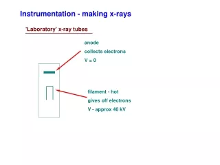
Instrumentation - making x-rays 'Laboratory' x-ray tubes anode collects electrons V = 0 filament - hot gives off electrons V - approx 40 kV
Instrumentation - making x-rays 'Laboratory' x-ray tubes anode collects electrons V = 0 L E K X filament - hot gives off electrons V - approx 40 kV = hc/E (Å) = 12.4/kV
Instrumentation - making x-rays 'Laboratory' x-ray tubes anode focal spot Filament geometry & electron focusing determine size, shape of focal spot, which influences size, shape of beam from tube
Instrumentation - making x-rays 'Laboratory' x-ray tubes anode focal spot Filament geometry & electron focusing determine size, shape of focal spot, which influences size, shape of beam from tube For highest intensity, want smallest size possible, but power limited by rate of heat transfer in anode
Instrumentation - the beam 'Laboratory' x-ray tubes anode focal spot 1st problem - tubes usually have liner focal spot w/ finite width microfocus tubes
Instrumentation - the beam 'Laboratory' x-ray tubes anode focal spot 1st problem - tubes usually have liner focal spot w/ finite width microfocus tubes 2nd problem - x-ray spectrum intensity wavelength
Instrumentation - the beam 'Laboratory' x-ray tubes anode focal spot 1st problem - tubes usually have liner focal spot w/ finite width microfocus tubes 2nd problem - x-ray spectrum Need beam conditioning intensity wavelength
Instrumentation - the beam 'Laboratory' x-ray tubes Pinhole collimator large intensity loss spectrum problems remain beam divergence probs looking at beam
Instrumentation - the beam 'Laboratory' x-ray tubes Pinhole collimator large intensity loss spectrum problems remain beam divergence probs Monchromator nearly single WL beam size probs yet beam divergence probs intensity wavelength
Instrumentation - the beam 'Laboratory' x-ray tubes Solns: Decrease focal spot size power limited, but flux still high spectrum probs remain
Instrumentation - the beam 'Laboratory' x-ray tubes Solns: Decrease focal spot size power limited, but flux still high spectrum probs remain Use new optics flux high monochromatic
Instrumentation - the beam 'Laboratory' x-ray tubes Use new optics flux high monochromatic
Instrumentation - the beam 'Laboratory' x-ray tubes Use new optics flux high monochromatic 1-D example:
Instrumentation - the beam 'Laboratory' x-ray tubes Use new optics flux high monochromatic 2-D:
Instrumentation - the beam 'Laboratory' x-ray tubes Small focal spot + new 2-D optics + pinholes + distance small beam low divergence monochromatic
Instrumentation - saxs system 'Laboratory' x-ray tubes Small focal spot + new 2-D optics + pinholes + distance small beam low divergence monochromatic