Understanding the Cell Cycle: Interphase and Mitosis Explained
90 likes | 200 Views
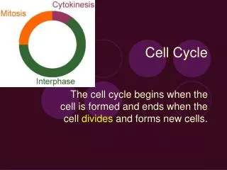
This guide explores the key phases of the cell cycle, focusing on interphase and mitosis. Interphase is the stage where the cell grows and replicates its DNA, often appearing to rest. Mitosis consists of four main phases: prophase, metaphase, anaphase, and telophase, where chromosomes become visible, align, separate, and the nuclear membrane reforms. Additionally, cytokinesis is the final step of cell division, resulting in two distinct daughter cells. Learn more about each phase and its significance in cell reproduction.

Understanding the Cell Cycle: Interphase and Mitosis Explained
E N D
Presentation Transcript
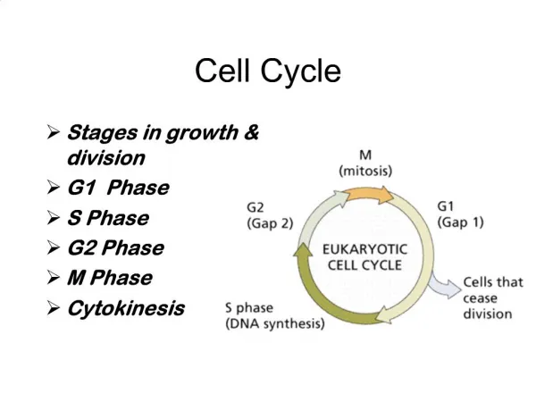
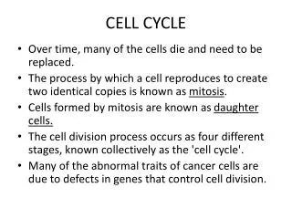
Cell cycle Shannon Abbott
Interphase fig2 Interphase is the beginning of the cell cycle. Interphase is where the cell is still growing. The DNA is replicating itself. It also is looking like it is resting. fig1 fig2http://staff.jccc.net/pdecell/celldivision/mitosis1.html fig1http://faculty.ccbcmd.edu/courses/bio141/lecguide/unit6/genetics/DNA/DNArep/pcinterphase.html
Mitosis The steps of mitosis: • prophase • Metaphase • Anaphase • Telophase
Prophase fig2 fig1 Mitosis Prophase: The chromosomes are finally becoming visible. The nuclear membrane is breaking down. The spindle fibers are appearing. fig2http://s37snpg.edu.glogster.com/portfolio/glog-book/ fig1http://faculty.ntcc.edu/mhearron/mitosis_and_meiosis.htm
Metaphase Fig 1. Fig 2. Mitosis metaphase: The chromosomes are lining up in the middle. The fibers are attach to the centromere of the chromosomes. fig1http://www2.sunysuffolk.edu/gambier/micrographs/metaphase.htm fig2http://faculty.clintoncc.suny.edu/faculty/Michael.Gregory/files/Bio%20100/Bio%20100%20Lectures/mitosis/mitosis.htm
Anaphase fig2 mitosis: anaphase The duplicated chromosomes separate. The spindles fibers pull the chromosomes away from each other. The cytoplasm division begins. fig1 fig1http://faculty.ccbcmd.edu/courses/bio141/lecguide/unit6/genetics/DNA/DNArep/acanaphase.html fig2http://www.sparknotes.com/biology/cellreproduction/mitosis/section2.rhtml
Telophase fig1 Mitosis: Telophase The nuclear membrane reforms. The chromosomes become less visible. The cytoplasm division is still occurring. http://www.edupic.net/cells.htm
Cytokinesis The cells are splitting in this stage. The cells are pinching and squeezing away from each other and finally spit away from each other. fig1 http://www.vtt.fi/uutta/2008/16092008_soluttuuliajolla.jsp?lang=en