COPD: Pathogenesis, Pathophysiology, and Physical Signs
Chronic Obstructive Pulmonary Disease (COPD) is a common respiratory condition characterized by chronic bronchitis and emphysema. This text explores the pathogenesis, pathophysiology, and physical signs of COPD, including the effects of smoking, air pollution, occupation, infections, and genetic factors. Learn about the two distinct processes involved, epidemiology, and classification of COPD. Gain insight into the pathology of chronic bronchitis and emphysema, and understand the physical signs that may indicate COPD.

COPD: Pathogenesis, Pathophysiology, and Physical Signs
E N D
Presentation Transcript
COPD SS Visser, Pulmonology Internal Medicine UP
Chronic Obstructive pulmonary Disease • Two distinct processes are involved, most often in combination. • Chronic Bronchitis – dx on history • Emphysema – dx previously on histology, nowadays clinically (good clinical-pathologic-radiologic correlation)
Def: Chronic Bronchitis • Excessive tracheobronchial mucus production sufficient to cause cough with expectoration for most days of at least 3 months of the year for 2 consecutive years. • Classification: • Simple chronic bronchitis • Chronic mucopurulent bronchitis • Chronic bronchitis with obstruction • Chronic bronchitis with obstruction and airway hyperreactivity.
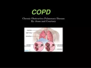
Def: Emphysema • Permanent abnormal distention of air spaces distal to the terminal bronchiole with destruction of alveolar septa (containing alveolar capillaries) and attachments to the bronchial walls. • Classification: • Centriacinar ( centrilobular) emphysema • Panacinar emphysema • Paraseptal emphysema • Senile emphysema
Def: COPD • Chronic obstruction to airflow due to chronic bronchitis and/or emphysema. • Degree of obstruction may be less when the patient is free from respiratory infection and may improve with bronchodilator drugs • Significant obstruction is always present
Epidemiology of COPD • 30% of smokers develop COPD • 20% of adult males have COPD • 15% of COPD patients are severely symptomatic • 4 th leading cause of death (USA) • Mortality rate still rising • prevalence in low birth weight and low socioeconomic status • Tuberculosis in smokers predisposes to COPD
Pathogenesis:Effects of Smoking -1 • Oxidative stress: O2-, OH-,H2O2, HOCl; source of Fe2+ catalizes production of OH- by neutrophils, eosinophils, alveolar macrophages; tar (cigarettes) contains NO and induces iNOStoxic peroxynitrites • Elastin breakdown- activated neutrophils neutrophil elastases and oxidants; -1-AT and metalloproteinase inhibitors (lung defenses) inactivated by smoke • Chemoattractant, upregulation of adhesion molecules neutrophil sequestration in lungs • expression of pro-inflammatory mediators: IL-8, NF-B recruitment of N, B, E and T lymphocytes
Effects of smoking -2 • levels of myeloperoxidase and eosinophilic cationic protein bronchoconstriction • levels of TGF- (transforming growth factor) fibrogenesis • Lipid peroxidation and DNA damage point mutations 0f the p53 gene locus epithelial dysplasia and lung cancer • ciliary function retained secretions; airway resistance vagal-mediated smooth muscle contraction • Hypertrophy and hyperplasia of mucus secreting glands secretions
Pathogenesis-3 • Air pollution exacerbations of CB related to heavy pollution with SO2 and NO2 • Occupation exposure to organic and inorganic dust or noxious gases accelerated decline in lung function • Infection even mild viral respiratory infections ( rhino virus) may be a major factor associated with etiology as well as progression of disease; severe viral pneumonia early in life may lead to COPD • Genetic factors: - -1-antitrypsin deficiency PIZZ, PISZ, PI00 (PI null null), susceptibility to effects of smoking
Pathophysiology • Air trapping- RV and FRC elevated • Hyperinflation –TLC elevated • elastic recoil pressure dynamic collapse of airways during expiration ineffective cough mechanism and pursed lips breathing (emphysema) • compliance (emphysema) • airway resistance • Prolonged forced expiratory time (N=<6 seconds)
Pathology: CB • Hypertrophy of mucus-producing glands in submucosa of large cartilaginous airways • Goblet cell hyperplasia, mucosal and submucosal inflammatory cell infiltrate, oedema, peribronchial fibrosis, intraluminal mucus plugs and increased smooth muscle in small airways • The major site of airflow obstruction is in the small airways and the inflammatory infiltrate consists of neutrophils (in asthma eosinophils)
Pathology : Emphysema • in number and size of alveolar fenestrae eventual destruction of alveolar septa and their attachments to terminal and respiratory bronchioles distention of alveolar spaces • Centriacinar E- respiratory bronchioles (central) affected • Panacinar E- central and peripheral portions of acinus affected • Senile E- alveoli and alveolar ducts enlarge (> 50 Y) • Periacinar/paraseptal E- distention of alveolar spaces adjacent to septal and pleural surfaces
Physical signs of COPD • Ronchi- in early disease present on forced expiration, later present in inspiration and expiration • Prolonged forced expiratory time (> 6 seconds) • Hyperinflation: cardiac dullness, liver dullness displaced downwards, A-P chest diameter, heart and breath sounds, Hoover sign • Inspiratory crepitations (lung bases) • Pursed lips breathing ( dynamic airway collapse) • Use accessory respiratory muscles • Signs of cor pulmonale and PHT
Emphysema = pink puffer Age (Dx) 60 + y Rest dyspnea mild-mod Exer dyspnea severe Cough ± Sputum scanty, mucoid Resp infect less often Resp failure terminal Cor pulmonale terminal Chronic Bronchitis = blue bloater 50 ± y none moderate prominent large volume, purulent often repeatedly common Emphysema:ChronicBronchitis
PHT (rest) 0-mild (exertion) moderate Build Asthenic, cachectic Hematocrit 35-45 Breath pattern use accessory muscles of respiration Sleep pattern Normal XRC Hyperinflation Bullae Mild-moderate severe obese, cyanosed 50-55 do not use accessory muscles of respiration sleep apnea bronchovascular markings Emphysema:Chronic Bronchitis
Blood gas: PaO2± 65 mm Hg PaCO2 35-40 Elastic recoil AW resistance N- Diffusion Cap FEV1 Bronchodilator response Poor 45-60 50-60 Normal N- Better but < 12% and 200ml Emphysema:Chronic Bronchitis
Spirometric classification of COPD severity using post-bronchodilator FeV1 • Stage I (Mild): FeV1/FVC <0.7; FeV1 80% of predicted • Stage II (Moderate): FeV1/FVC <0.7; FeV1 50- <80% of predicted • Stage III (Severe): FeV1/FVC <0.7; FeV1 30-<50% • Stage IV (Very severe): FeV1/FVC <0.7; FeV1 <30% or <50% but chronic respiratory failure is present. (GOLD 2007)
Treatment: Goals of management -1 • Recognition of disease (early Diagnosis and staging) • Smoking cessation (secondary prevention) nicotine replacement and Zyban • Improvement of breathlessness (Rx of airflow obstruction- bronchodilator drugs) 1.Methylxanthines 2.Short and long-acting B2adrenergic agonists ( incidence of pneumonia with ICS and LABA combinations) 3.Short and long-acting Anticholinergics- BD of choice in COPD
Treatment -2 • Respiratory infections –AB when sputum volume and/or purulence (exacerbation of COPD); Influenza and Streptococcus pneumoniae vaccination • Bronchopulmonary drainage and postural drainage (physiotherapy) for patients with CB • Oxygen therapy for patients with hypoxia (PaO2<55 mmHg, SaO2 <88% ) and erythrocytosis (Hematocrit>55) • Pulmonary rehabilitation and education ( improving quality of life)- exercise program and improved nutrition • Prevention and treatment of complications (cor pulmonale) and limitation of disease progression
Treatment -3 • Glucocorticoids –only 10% of COPD patients show subjective benefit and improved lung function (FeV1 increase of 20% or more) on systemic GCs; with COPD exacerbation a course of prednisone 40 mg/d for 2 weeks are usually prescribed • Inhaled GCs may severity of exacerbations and need for hospitalisation. Benefit of 10-14 day trial of 30-40mg prednisone for Stage III COPD patients remains to be proven. • Lung volume reduction surgery • Transplantation
Airway Diseases - COPD • Smoking • Hyperinflation • Airway collapse • Respiratory infection • Bronchospasm • Allergy • Inflammation
Airway Diseases : Asthma • Allergy • Inflammation • Bronchospasm • Hyperinflation • Respiratory infection
AirwayDiseases:Bronchiectasis • Respiratory infection • Hyperinflation • Bronchospasm • Inflammation • Allergy