Download
1 / 38
380 likes | 493 Views
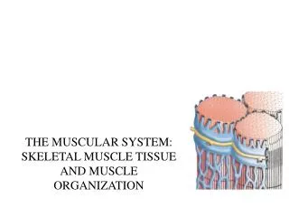
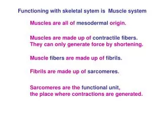
This overview covers the key characteristics of muscles, including irritability, contractility, extensibility, and elasticity. It discusses muscle functions such as movement, posture, and heat production, highlighting the roles of skeletal, smooth, and cardiac muscles. The structure of skeletal muscle, including the epimysium, perimysium, and endomysium, is detailed, along with the anatomy of muscle cells, such as myofibrils and myofilaments. This comprehensive guide is essential for grasping muscle function and structure in human physiology.

E N D
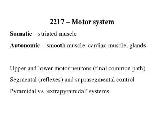
Characteristics of Muscles • Irritability (excitability) • Respond to chemicals (neurotransmitters), stretch etc. by altering their plasma membrane electrical charge • Contractility • Can shorten their length by a substantial amount when stimulated • Extensibility • Can stretch back to their original length after each contraction • Elasticity • When stretched and then released, muscle will recoil to its original resting length
Muscle Functions • Movement • Body movement and mobility and various bodily functions • Posture and body support • Maintain body position • Heat production • 85% body heat is generated by muscle contractions
Muscle types • Skeletal muscle • Voluntary • Striated • Usually attached to bone • Smooth muscle • “fusiform” cells • Involuntary • “Pacemaker” cells usually spontaneously depolarize and stimulate contractions (autorhythmic) • Autonomic nervous system can stimulate contractions as well • Cardiac muscle (heart muscle) • Involuntary • “pacemaker” cells autorhythmic • Autonomic nervous system control • Striated, like skeletal muscle cells • Remember the intercalated discs?
Skeletal muscle - Attachments • Usually attached to 2 or more bones (span a joint) • Voluntary: under voluntary/conscious control • Skeletal muscle cells = “muscle fibers” or “myofibers” • Range from 3-30 cm long • Attached to the bone by tendons (in most cases)
Skeletal muscle – Attachments • “sheaths” around muscle fibers • Endomysium: areolar connective tissue • Where blood capillaries, neural inputs are located • Perimysium: bundles a number of muscle cells together • The bundles of muscle cells = “fascicles” • Epimysium: surrounds the entire muscle • Deep fasciae: separates adjacent muscles • Devoid of adipose cells. • Superficial fasciae (hypodermis): between muscle and dermis (skin) • Very adipose-cell dense in some areas (abdomen & buttocks • Note: The structures attaching the muscle are extension of the connective tissue forming the endo/peri/epimysium
Muscle structures - Attachments • Deep fascia: fibrous connective tissue anchoring groups of muscle and deep structures, support nerves, blood vessels, forms tendons, ligaments, joint capsules. • Superficial fasciae: support the skin, nerves and blood vessels, provides elasticity to hypodermis • Aponeuroses: sheet-like tendons- ex: over the skull • Tendon sheath: encloses group of thin tendon in a sheath, for protection and lubrication– wrist. • Retinaculum:sheat of connective tissue binding a group of tendons
Aponeuroses in scalp and abdomen Retinaculum in your wrist
Applications • Compartment syndrome - due to muscle swelling In order to prevent limb loss, the tissues are slashed
Muscle attachment • Epimysium: surrounds the entire muscle • Recall that tendons usually attach to bone via the periosteum (outer membrane on a bone) • 2 types of muscle-to-bone connections: • “direct” or “fleshy attachment” • Collagen fibers of epimysium continue in the periosteum • Looks like the muscle is directly attached to bone (in your ribs) • “indirect” attachment • Epimysium is in unison with tendon • The tendon attaches to bone • Seen as a white or pale “gap” between the red part of a muscle and the bone
Skeletal muscle cells • Derived from stem cells called “myoblasts” • Several myoblasts will fuse together during embryonic development • Reason why skeletal muscle cells are multinucleate (have many nuclei)
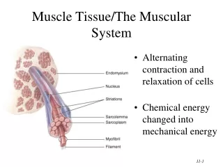
Skeletal muscle cells • Muscle cell (myocyte, muscle fiber) anatomy: • Outer plasma membrane = sarcolemma (“flesh-lining”) • Sarcoplasm: cytoplasm of the myocyte • Myofibrils: Bundles of protein running along the length of the cell • Myoglobin: similar to hemoglobin • Stores oxygen within the muscle cell • Important for ATP synthesis during contraction
Skeletal muscle cells • Muscle cell (myocyte, muscle fiber) anatomy: • Sarcoplasmic reticulum: specialized area of the endoplasmic reticulum • Surrounds the myofibrils like a web • At each end, forms bag-like structure “terminal cisternae” • Area where calcium (Ca2+) is stored • Transverse tubules: tube formation of the sarcolemma • Tubes run from the external surface straight through the muscle cell • Transverse tubules and terminal cisternae form a “triad” structure (many of these along the length of the muscle cell)
Transverse tubules form a “pore” through which the muscle cell can “sense” it’s outside environment and respond to stimuli
Myofibrils • Made of “myofilaments”: • Thick myofilaments: bundles of myosin • Thin myofilament: Actin
Myosin • Myosin head on the thick filament will try to bind to the actin bead at a specific “active” site on the actin molecule • Also has “control” proteins to control contraction (control myosin attachment)
Actin • Thin myofilaments: • Globular actin filamentous actin • Tropomyosin: “plugs” the site where myosin wants to attach • Troponin: calcium-sensitive protein
The sarcomere • “sarcomere”: refers to the entire complex between each Z-disc. • Formation of bands: • - A bands: Myosin + actin myofilaments • - I band: actin only
In summary http://www.life.uiuc.edu/crofts/bioph354/lect16&17.html
Motor unit • All muscle fibers controlled by 1the same motor neuron • Which of the two set up provides the finest muscle control? • 10 muscle fibers controlled by 1 motor neuron • Or • 1000 muscle fibers controlled by 1 motor neuron
Contraction • During contraction, the myosin “rows” along the actin and pull the Z-discs closer to one another • The I band shortens • The A band do not change • http://www.blackwellpublishing.com/matthews/myosin.html • http://www.ebsa.org/npbsn41/intro_muscle.html
Skeletal muscle terms • “origin” = area where muscle attaches to bone that does not move (remains stationary) • “belly” = thick region of the muscle • “insertion” = area where muscle attaches to bone that moves
Skeletal muscle anatomy • Different forms of skeletal muscle: • Fusiform: thick in the middle, thin at ends • Biceps branchii, gastrocnemius in calf • Parallel: long, strap-like • Spans great lengths, can shorten/contract more than other muscles • Weaker than fusiform muscles • Rectus abdominus (abs…6-pack) • Convergent: fan shaped/leaf shaped • Large middle that tapers or focuses on 1 narrow insertion • Pectoralis major in the chest • Pennate and circular muscles (next slide)
Skeletal muscle anatomy • Pennate muscles: feather shaped - The muscle fibers attach obliquely or at an angle to the tendon • Unipennate: all muscles approach on 1 side of the tendon • Palmar interosseus of the hand • Bipennate: muscle fibers attach on both sides of the tendon • Rectus femorus of the thigh • Multipennate: a branched tendon, with muscle fibers attaching throughout • Deltoid of the shoulder • Circular muscle: sphincters etc. - Forms a ring around an opening/passage • Orbicularis oculi around the eye • Anal sphincter
Skeletal muscle anatomy • Each muscle requires nutrients & oxygen • Each muscle usually has at least 1 artery entering • Larger muscles (quadriceps femoris etc.) have 2+ arteries to bring blood etc. • Contraction requires a great deal of skeletal muscle cell metabolic activity
Skeletal muscle terms • Insertion & origin will differ depending on the MOTION • Quadriceps muscle: • When you kick (extend your knee), the quadriceps moves the tibia • Tibia is the insertion (moves), and the femur is the origin (stays still) • When you sit down, the terminology changes: • Tibia stays still (origin), and the femur moves (insertion)
Skeletal muscle terminology • Common muscle terms: • Shape terms (rhomboid, trapezius) • Location (pectoralis, intercostal, brachii, femoris) • Attachment (zygomaticus, temporalis, sternocleidomastoid) • Size (maximus, medius, minimus, longus, brevis) • Fiber orientation • Rectus = straight fibers (rectus abdominus) • Transverse = across (transverse abdominal muscle) • Oblique = slant/angle (external oblique muscle) • Relative position (lateral, medial, internal, external) • Function (adductor, abductor, extensor, flexor, pronator, levator)
Skeletal muscle movements • “action” = movement produced by a muscle • Note that skeletal muscle rarely acts independently or alone…almost always acts in groups • 4 major muscle action categories: • Prime mover (agonist) • Muscle that produces the most force during a particular joint movement • When you flex your elbow, your prime mover is the biceps brachii
Skeletal muscle movements • 4 major muscle action categories: • Prime mover (agonist) • Synergist • Muscle (s) that aid the prime mover • Usually act to stabilize the joint…focus the energy of the prime mover efficiently • Antagonist • Opposes the prime mover (muscles usually act in pairs…prime mover and antagonist) • Fixator • Prevents inappropriate movement • When you flex your elbow, you use the biceps brachii
Diseases of myofilaments • Muscular dystrophy: mutations in the dystrophin gene • Different types: • Duchenne dystrophy • Becker dystrophy
Myasthenia gravis Autoimmune disease: antibodies destroy the Acetylcholine receptors muscle weakness
Tetanus Due to a bacterial toxin http://medinfo.ufl.edu/year2/mmid/bms5300/images/a5.jpg http://www.humanillnesses.com/original/images/hdc_0001_0003_0_img0264.jpg www.4to40.com/images/qa/tetanus.jpg
Types of muscle contractions • Isometric contraction: contraction without a change in muscle length • When you hold in one place…it takes muscles to hold your book in front of you, even if none of the muscles in your arm are changing length • Isotonic contraction: contraction resulting in a change in muscle length, but no alteration in muscle tension • When you increase the internal tension in the muscle to overcome the resistance
Aging and muscle • Varies with people • Decrease in muscle mass • Very dependent on the person’s level of exercise • “If you don’t use it, you loose it”
Muscle imbalances • Eye muscles: various diseases, ex: strabismus • Torticolis