Download
1 / 30
320 likes | 414 Views
Learn about the skeletal and muscle systems, the types of muscles, how muscles work, muscle naming conventions, and common muscle disorders such as Muscular Dystrophy.

E N D
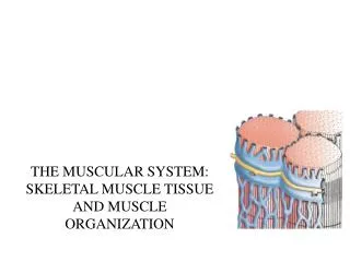
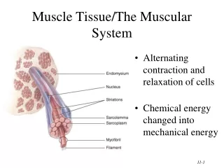
Functioning with skeletal sytem is Muscle system Muscles are all of mesodermal origin. Muscles are made up of contractile fibers. They can only generate force by shortening. Muscle fibers are made up of fibrils. Fibrils are made up of sarcomeres. Sarcomeres are the functional unit, the place where contractions are generated.
Muscle types innervation location fiber type fiber arrangement Smooth muscle Smooth parallel autonomic viscera Skeletal muscle parallel voluntary Striated Striated muscle Inter- connected autonomic heart Striated Cardiac muscle
Muscles Muscles are made up of fibers. Fibers are made up of fibrils (these are the cells). Fibrils are made up of sarcomeres. Z lines between sarcomeres produce striations in striated muscle.
Each sarcomere is made up of: BLUE RED
Simplified model. One sarcomere. Actin filaments. Mysoin filaments. During contraction, mysoin filaments latch on to adjacent actin filaments via myosinheads. The heads change shape pulling the actin filaments towards one another.
actin myosine
3 4 2 1
Skeletal Muscles Contractile fibers make up belly of muscle Synovial joint Only work by getting shorter, by contracting Belly of muscle Force of contraction transmitted by tendons (in yellow) Tendons do not contract tendon Force results in movement, usually of bones at joints Synovial joint
Skeletal Muscles All have an origin. Synovial joint Site of attachment closer to the midline All have an insertion Belly of muscle Site of attachment away from the midline These are standard descriptors of any muscle. tendon They are often used in a muscle name. Synovial joint e.g. coracomandiularis
Because muscles can only work by contraction they must work in antagonistic pairs. Antagonistic pairs have opposite actions. In this case: Flexion (biceps) Extension (triceps) The names of muscles frequently use the name of the action of the muscle. e.g. Flexor digitorum profundus
Learning the names of muscles. The names always describe properties of the muscle, such as: 1) origin and insertion. coracomandiularis xiphihumeralis sternomastoid cleidomastoid 2) number of heads or origins biceps triceps 2) depth of the muscle pectoralis profundus flexor digitorum superficialis
Learning the names of muscles. The names always describe properties of the muscle, such as: 4) relative position of the muscle. flexor carpi ulnaris flexor carpi radialis lateral or medial head of triceps 5) length of a tendon Adductor pollicus longus Adductor pollicus brevis 6) action of the muscle Flexor digitorum profundus Extensor digitorum communis
6) The actions of skeletal muscles Always occur in pairs Actions will be oppsite of each other extensor carpi ulnaris Extension -- make an angle larger flexor carpi ulnaris Flexion -- make an angle smaller adduction -- pull towards midline fin adductor abduction -- pull away from midline fin abductor protraction -- reach forward from midline tibialis cranialis gastrocnemis retraction -- pull back towards midline pronation -- rotate thumb away from midline pronator supinator supination -- rotate thumb towards midline
Muscles function in lever systems Fi x Li = Fo x Lo There is more to the system than mechanical advantage
But there is more than mechanical advantage Distal end of arm (hand) moves much faster and much farther than proximal end. L Allows for acceleration and wider range of movement.
Muscular Dystrophy Kaitlyn Gilligan
Muscular Dystrophy- is a genetic disorder that weakens the muscles that help the body move. People with MD have incorrect or missing information in their genes, which prevents them from making the proteins they need for healthy muscles.
Types of MD • Becker • Congenital • Distal • Duchenne • Emery-Dreifuss • Facioscapulohumeral • Limb-girdle • Myotonic • Oculophayngeal The symptoms of muscular dystrophy are the result of the deterioration of the body’s muscles. Due to the death of muscle cells and muscle tissues it leads to ongoing muscle wasting and muscle weakness. MD progresses and gets worse over time.
Duchenne Muscular Dystrophy- most common form of MD among children. DMD occurs in approximately 1 in 3,500 to 6,000 male births. Usually a person loses the ability to walk between 7-13 years old. Myotonic Dystrophy- a form of MD when the muscles have difficulty relaxing . It can cause muscle weakness and wasting, cataracts, and heart problems. DBMD- caused by a mutation in the gene that makes dytrophin. Dystrophin helps muscles stay healthy and strong. Boys with DBMD have little or no dystrophin. Becker Muscular Dystrophy- milder form of MD. Affects about 1 in 18,500 males.
Symptoms/Signs Signs of DBMD • Experience a delay in walking • Having large calf muscles • Falling more often than children • Walking on tip toes • Signs of Duchenne • Fatigue • Learning difficulties • Difficulty with motor skills • Progressive difficulty walking • Breathing difficulties • Signs of Myotonic • Difficulty swallowing • Gagging • Stiff movements that improve when they are repeated.