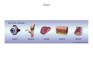
Senses
Senses. Sensation: -response to environment via generation of nerve impulse - sensation occurs upon arrival of nerve impulse at cerebral cortex and processing - perception is the interpretation of these sensations after processing

Senses
E N D
Presentation Transcript
Senses Sensation: -response to environment via generation of nerve impulse -sensation occurs upon arrival of nerve impulse at cerebral cortex and processing -perception is the interpretation of these sensations after processing -nerve impulses from the body sent via ascending tracts in spinal cord to the brain -from the special senses – through the brain stem
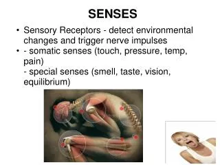
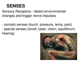
Senses • can be classified in several ways • based on: • 1. what they respond to • 1. Chemoreceptors • 2. Mechanoreceptors • 3. Nociceptors/pain receptors • 4. Thermoreceptors • 5. Photoreceptors • 2. how they are structured • e.g. corpuscles, free nerve endings • 3. their location • e.g. exteroceptors, interoceptors, proprioceptors, cutaneous receptors
Proprioceptors -located in muscles, joints and tendons -position of limbs and degree of muscle relaxation -proprioceptor: sensory neuron wrapped around a muscle fiber (muscle spindle), found embedded in a tendon or found in a joint -stretching of muscle fiber, tendon or flexing of joint stimulates the receptor Patellar reflex: -muscle stretch, proprioceptor fires impulse to spinal cord, reflex arc results, muscle fiber response
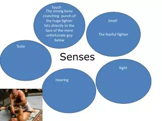
General vs. Special Senses • General • Touch, pressure, itch, tickle, heat/cold = cutaneous receptors (touch) • Proprioception - proprioceptors • Special • Sight • Hearing • Vision • Smell
Cutaneous receptors – Sensation of Touch -located in skin -receptors can be classified as cutaneous, or thermoreceptors/mechanoceptors etc…. -impulse sent to somatosensory areas of brain -touch receptors: Meissner’s (fingertips, lips, tongue, nipples, penis/clitoris) Merkel disks (epidermis/dermis) Root hair plexus (root of hair) -pressure receptors: Pacinian corpuscles -temp receptors: free nerve endings that respond to cold OR warmth -Krause end bulbs, Ruffini endings
Taste -taste requires dissolving of substances in saliva -taste buds: salty, sweet, bitter and sour – more???? -10,000 taste buds found on tongue, soft palate & larynx -one taste bud responds to one king of tastant -many clustered at specific locations – e.g. tip of tongue = sweet taste buds -found associated with projections called papillae -three types of papilla will bear taste buds -buds open at a taste pore which opens into a canal which opens onto the surface of the tongue salty bitter sour • taste papillae: • foliate • fungiform • circumvallate • filliform (texture) – no taste buds
Anatomy of Taste Buds • An oval body consisting of 50 receptor cells (gustatory or taste cells) surrounded by supporting cells • each taste cell projects a single gustatory hair projects upward through the taste pore • Taste pore exposed to saliva flowing over tongue • Basal cells develop into new receptor cells every 10 days.
Physiology of Taste • Complete adaptation in 1 to 5 minutes • Thresholds for tastes vary among the 4 primary tastes • most sensitive to bitter (poisons) • least sensitive to salty and sweet • Mechanism • dissolved substance contacts gustatory hairs – action potential • action potential results in neurotransmitter release and stimulation of the 1st order neuron synapsing with the gustatory neuron • 1st order neuron forms cranial nerves, VII, IX and X
Gustatory Pathway • gustatory fibers/axons found in three cranial nerves • VII (facial) serves anterior 2/3 of tongue • IX (glossopharyngeal) serves posterior 1/3 of tongue • X (vagus) serves palate & epiglottis • Signals travel to thalamus - extend from the thalamus to the primary gustatory area on parietal lobe of the cerebral cortex • provide conscious perception of taste • Taste fibers also extend to limbic system – association of taste with a memory
Smell • -requires dissolving of chemicals in mucus • -neurons = olfactory cells - located within olfactory epithelium in the nasal cavity • -olfactory epithelium covers superior nasal cavity (superior nasal conchae) • and cribriform plate • -contains neurons (olfactory cells) and supportive cells and basal cells
Olfactory epithelium • Olfactory receptors • bipolar neurons with cilia or olfactory hairs • Supporting cells • columnar epithelium • Basal cells = stem cells • replace receptors monthly • Olfactory glands • produce mucus • Both epithelium & glands innervated by cranial nerve VII.
Eustacian tube With tubal tonsil
Olfaction: Sense of Smell • Odorants bind to receptors • Na+ channels open • Depolarization occurs • Nerve impulse is triggered • each olfactory neuron can respond to multiple odorants
Olfactory Pathway • Olfactory neurons pass through the foramina in the cribriform plate • Synapse with the neurons of the olfactory bulb • Axons of these neurons form the olfactory nerves (Cranial nerve I) • axons within the olfactory bulb form the olfactory tract • synapses on the primary olfactory area of temporal lobe • conscious awareness of smell begins • Other pathways lead to the frontal lobe (Brodmann area 11) where identification of the odor occurs
Adaptation & Odor Thresholds • Adaptation = decreasing sensitivity • Olfactory adaptation is rapid • 50% in 1 second • complete in 1 minute • Low threshold • only a few molecules need to be present • e.g. methyl mercaptan added to natural gas as warning
Vision -sight is generated by the bending and focusing of light onto the retina • Anterior cavity (anterior to lens) • filled with a liquid aqueous humor • produced by ciliary body • continually drained • replaced every 90 minutes • 2 chambers • anterior chamber between cornea and iris • posterior chamber between iris and lens • Posterior cavity (posterior to lens) • filled with vitreous body (jellylike) • formed once during embryonic life
Accessory Structures of Eye • Eyelids or palpebrae • protect & lubricate • epidermis, dermis, CT, orbicularis oculi m., tarsal plate, tarsal glands & conjunctiva • Tarsal glands • oily secretions keep lids from sticking together • Conjunctiva • palpebral & bulbar • stops at corneal edge • dilated BV--bloodshot
Extraocular Muscles • Six muscles that insert on the exterior surface of the eyeball • Innervated by CN III, IV or VI. • 4 rectus muscles -- superior, inferior, lateral and medial • 2 oblique muscles -- inferior and superior
Lacrimal Apparatus • About 1 ml of tears produced per day. Spread over eye by blinking. Contains bactericidal enzyme called lysozyme.
Tunics (Layers) of Eyeball • Fibrous Tunic(outer layer) • Sclera and cornea • Vascular Tunic(middle layer) • Choroid • Iris • Ciliary body • lens • Nervous Tunic(inner layer) • retina
Fibrous Tunic • CORNEA • Transparent • Helps focus light (refraction) • Astigmatism = uneven cornea • 3 layers • nonkeratinized stratified squamous (outer) • collagen fibers & fibroblasts • simple squamous epithelium • Nourished by tears & aqueous humor • SCLERA • “White” of the eye • Dense irregular connective tissue layer -- collagen & fibroblasts • Provides shape & support, attachment for eye muscles • Posteriorly pierced by Optic Nerve (CNII)
Vascular Tunic • Choroid • Majority of vascular layer • Found underneath the retina • pigmented epithelial cells that make melanin (melanocytes) & blood vessels • provides nutrients to retina • black pigment in melanocytes absorb scattered light and ensure light passes through retina • Lens: • Avascular • Crystallin proteins arranged like layers in onion • Clear capsule & perfectly transparent • Lens held in place by suspensory ligaments which attach to the ciliary processes • Focuses light on fovea (center of the retina)
Vascular Tunic • Ciliary body • choroid extends to the front of the eye as ciliary musclesand ciliary processes – for controlling the shape of the lens • ciliary processes • folds on ciliary body • secrete aqueous humor • ciliary muscle • smooth muscle that alters shape of lens • attach to the ciliary processes • Suspensory ligaments attach lens to ciliary process • Ciliary muscle controls tension on ligaments & lens
Vascular Tunic Aqueous Humor • Continuously produced by ciliary body • Flows from posterior chamberinto anterior through the pupil • Scleral venous sinus • canal of Schlemm • opening in white of eyeat junction of cornea & sclera • drainage of aqueous humor from eye to bloodstream
Vascular Tunic -- The Iris • is a coloured extension off the ciliary processes • Constrictor pupillae muscles (circular muscles) • are innervated by parasympathetic fibers while • Dilator pupillae muscles (radial muscles) • are innervated by sympathetic fibers • Contraction results in pupil dilation • Response varies with different levels of light Constriction of the Pupil • Constrictor pupillae muscle contracts • Narrows beam of light that enters the eye • Prevents light rays from entering the eye through the • edge of the lens • Sharpens vision by preventing blurry edges • Protects retina very excessively bright light
Nervous Tunic Retina • Posterior 3/4 of eyeball • anterior edge = ora serrata • Optic disc • optic nerve exiting back of eyeball • attachment of retina to optic nerve - optic disc (blind spot) • central depression in retina - fovea centralis View with Ophthalmoscope
Layers of Retina • Pigmented epithelium • nonvisual portion • absorbs stray light & helps keep image clear • 3 layers of neurons • photoreceptor layer • bipolar neuron layer • ganglion neuron layer • 2 other cell types (modify the signal) • horizontal cells • amacrine cells CHOROID Posterior cavity
Photoreceptors -rod and cones -rod cells: black and white, bright and dark -cone cells: colour vision -visual pigment:rhodopsin:opsin and retinal -visual pigment is folded into “discs” in the outer segment of the photoreceptor -inner segment - cell body -synaptic endings for neurotransmitter release • Rods----rod shaped outer segment • shades of gray in dim light • 120 million rod cells over the retina • discriminates shapes & movements • distributed along periphery for peripheral vision
Cones----cone shaped • sharp, color vision • 6 million total over the retina • 3 types: blue, red and yellow/green colour (differences in opsin structure) • Large concentration found in the • fovea of macula lutea (fovea centralis) • densely packed region of cones • at exact visual axis of eye • sharpest resolution or acuity • sharpest colour vision
Physiology of Vision • In darkness • Na+ channels are held open and photoreceptor is always partially depolarized - continuous release of an inhibitory neurotransmitter onto bipolar cells • The bipolar cells are prevented from creating their action potential • No nerve impulse leaves the retina = NO IMAGE • In light • Light is absorbed by the rhodopsin pigment - activates enzymes in the photoreceptor • These enzymes cause the closing of Na+ channels – hyperpolarizing the photoreceptor – no action potential!! • release of inhibitory neurotransmitter is stopped • bipolar cells can now spontaneously generate their action potential • this creates an action potential in the ganglion cells – via an electrical synapse • a nerve impulse will travel towards the brain = IMAGE
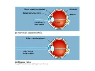
Major Processes of Image Formation • Refraction of light • light rays must fall upon the retina – so they must be bent • Refraction = Bending of light as it passes from one substance (air) into a 2nd substance with a different density (cornea) • In the eye, light is refracted by the anterior & posterior surfaces of the cornea and the lens • Accommodation of the lens • changing shape of lens so that light is focused • Constriction of the pupil • less light enters the eye
Refraction by the Cornea & Lens • Image focused on retina is inverted & reversed from left to right • Brain learns to work with that information – switches it back in the occipital lobe • For objects far away - 75% of Refraction is done by cornea -- rest is done by the lens • Light rays from more than 20’ are nearly parallel and only need to be bent enough to focus on retina – mainly by the cornea • Light rays from less than 6’ are more divergent & need more refraction – cornea and lens
Emmetropic eye (normal) • can refract light from 20 ft away • Myopia (nearsighted) • eyeball is too long from front to back • glasses concave • Hypermetropic (farsighted) • eyeball is too short • glasses convex (coke-bottle) • Astigmatism • corneal surface wavy • parts of image out of focus
Accommodation & the Lens • View a distant object • lens is nearly flat by pulling of suspensory ligaments • This requires relaxation of the ciliary muscles • View a close object • ciliary muscle is contracted & decreases the pull on the suspensory ligaments on the lens • elastic lens thickens as the tension is removed from it • this causes a rounding of the lens – more refraction • this extra process needed to get additional bending of light is called accommodation
Hearing & Equilibrium -outer ear:pinna - cartilage and skin -for collection of sound waves -middle ear: tympanic membrane and 3 ossicles (malleus, incus, stapes) -transmission of sound waves to inner ear -inner ear: cochlea (hearing), saccule, utricle & three semicircular canals (balance)
External Ear • Function = collect sounds • Structures • auricle or pinna • elastic cartilage covered with skin • external auditory canal • curved 1” tube of cartilage & bone leading into temporal bone • ceruminous glands produce cerumen = ear wax • tympanic membrane or eardrum • epidermis, collagen & elastic fibers, simple cuboidal epith. • Perforated eardrum (hole is present) • at time of injury (pain, ringing, hearing loss, dizziness) • caused by explosion, scuba diving, or ear infection
Middle Ear Cavity • Air filled cavity in the temporal bone • Separated from external ear by eardrum and from internal ear by oval & round window • These windows have membranes similar to the tympanic membrane • Will vibrate with the same frequency as the tympanic membrane • As the oval window pushes in the round window must bulge outwards to keep the pressure within the ear in a safe zone • 3 ear ossicles connected by synovial joints • malleus attached to eardrum, incus & stapes attached to the membrane of oval window • stapedius and tensor tympani muscles attach to ossicles – prevent large vibrations that would damage the inner ear • Auditory tube/Eustacian tube leads to nasopharynx • helps to equalize pressure on both sides of eardrum – better vibration and better sound
Inner Ear---Bony & Membranous Labyrinth • Bony labyrinth = set of tubelike bones carved into the temporal bone • surrounds & protects Membranous Labyrinth • filled with a fluid called perilymph • Membranous labyrinth = set of membranous tubes within the bony labyrinth • Filled with a fluid called endolymph • contains sensory receptors for hearing & balance and filled with endolymph • utricle, saccule, ampulla, 3 semicircular ducts & cochlea
Cochlear Anatomy • 3 fluid filled channels found within the cochlea • scala vestibuli, scala tympani and scala media (cochlear duct) • Vibration of the stapes upon the oval window pushes it in - sends vibrations into the perilymph of the scala vestibuli and tympani • The movement of the perilymph within these scala pushes into the cochlear duct and stimulates neurons within it • Fluid vibration dissipated at round window which bulges out as the oval window pushes in
Partitions that separate the channels are Y shaped • vestibular membrane above & basilar membrane below form a central fluid filled chamber = scala media (cochlear duct)
within the cochlear duct • organ of hearing = Organ of Corti • Organ of Corti– Tectorial membrane and a Basilar membrane surrounding sensory neurons • Neurons are also called hair cells –bear large cilia called stereocilia • project from the basilar membrane and the cilia are covered with the tectorial membrane • endolymph flowing through the cochlear duct shifts the basilar membrane back and forth • bends the hair cells, results in an action potential – potentials transmitted to sensory neurons within the 8th cranial nerve (acoustic or vestibulocochlear nerve)
Physiology of Hearing • #1: Auricle collects sound waves • #2: Eardrum vibrates • #3: Ossicles vibrate since malleus attached to eardrum • #4: Stapes pushes on oval window producing fluid pressure waves in scala vestibuli & tympani • #5 to #7: Waves push against the cochlear duct • Pressure fluctuations in the cochlear duct moves the basilar membrane back and forth • This moves the hair cells against the tectorial membrane and bends the stereocilia • Bending produces action potentials • #8: pressure of the fluid moving through the cochlea is dissipated as the round window bulges out • Cochlear branch of CN VIII gathers signals from neurons in cochlear duct • Sends signals to medulla oblongata (cochlear and superior olivary nuclei) • Nerves take this impulse from the MO and transmit them (via thalamus) to the primary auditory cortex in the temporal lobe
Physiology of Equilibrium (Balance) • Static equilibrium • maintain the position of the body (head) relative to the force of gravity • macula receptors within saccule & utricle • Dynamic equilibrium • maintain body position (head) during sudden movement of any type--rotation, deceleration or acceleration • crista receptors within ampulla of semicircular ducts
Static equilibrium: Saccule & Utricle • Thickened regions called macula within the saccule & utricle • Cell types in the macula region • hair cells (neurons with stereocilia) • supporting cells that secrete gelatinous otolithic • Gelatinous otolithic membrane contains calcium carbonate crystals called otoliths that move when you tip your head • head movement and otolith movement bends the hair cells and results in action potentials
Dynamic equilibrium: Semicircular Ducts • Small elevation within each of three semicircular ducts • anterior, posterior & horizontal ducts detect different movements • Hair cells covered with cupulaof gelatinous material • When you move, fluid in canal bends cupula stimulating hair cells that release neurotransmitter • This activates the neurons whose axons make up the vestibulocochlear nerve (VIII) • Signals run from VIII to cerebellum and to the medulla • Onto the temporal lobe and motor cortex