Download
1 / 10
140 likes | 351 Views
Explore the world of nanoscale imaging with our UV Photo-Emission Electron Microscopy system, offering real-time, high-resolution imaging capabilities down to <10 nm resolution. This system allows for the measurement of work function contrast, variable energy yields, and local work function extraction. With tunable synchrotron X-rays and UV light, and a sampling depth of 3-10 nm, this instrument enables full-field imaging and local measurements with exceptional precision. Coupled with pump-probe studies, it offers temporal resolution of ~15 fs for tracking plasmon wavepacket dynamics and surface interactions.

E N D
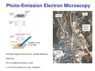
Photo-Emission Electron Microscopy CCD X-rays Electron Optical Column Tunable x-rays or UV light Transfer • Full field imaging of electrons (ie. parallel detection) • Real time • UV or tunable synchrotron x-rays • < 5 nm (UV) and 25 nm (x-ray) resolution
Electron Yield Sampling Depth 3-10 nm
UV Photo-Emission Electron Microscopy • UV imaging • work function contrast • < 10 nm resolution • variable energy • measure of yield (energy) • extract local work function 266 nm image of LCLS copper cathode, 30 x 30 microns
UV Photo-Emission Electron Microscopy p: 266 nm light PEEM image 7:1 hole to background contrast s: 266 nm light PEEM image 1.5:1 hole to background contrast • local measurement of field strength • local measurement of Fowler curves….work function • image as function of photon energy to 1 eV above work function 50 nm holes on 248 nm pitch, Al (FIB: Hyuck Choo)
Synchrotron PEEM: Elemental Contrast from Jo Stohr
Synchrotron PEEM: Chemical / Magnetic Contrast from Jo Stohr
A FM Co E p s [010] 45° 5 mm c c LaFeO3 AFM [100] Magnetic and Antiferromagnetic Domains In Co-LaFeO3 Scholl et al., Science, 2000 LaFeO3 Nolting et al., Nature 2000
100 psec pump probe PEEM magnetic domains Gradient image Cho et al., Science 2004
Image of electron yield as a function of time • temporal resolution ~ 15 fsec • Image shows propagation of the plasmon wavepacket + dispersion + damping • image surface using PEEM; Ag polycrystalline film • pump - probe geometry using 100 MHz laser oscillator, 10 fsec, 3 eV photons • launch SPP, then 2 photon photoemission image at delay dT Apply to plasmonic nanostructures (coupled to photovoltaic materials)
Photo-Electron Emission Microscopy • - Full field….50 microns • - 5 - 20 MV/m • - Secondary electron imaging • - Variable UV photon energy…..local Fowler plots……local work function • - Variable SR x-rays….local elemental and chemical speciation • - 10 nm resolution with UV, 25 nm with x-rays • - can be combined with pump probe time resolved studies