
Parasitology Laboratory Staining Techniques: Cryptosporidium parvum & Isospora belli
E N D
Presentation Transcript
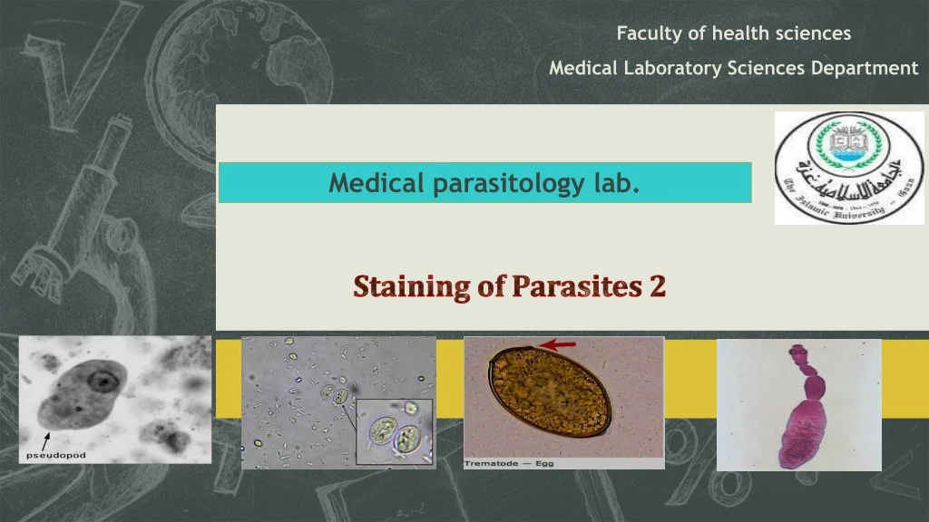
Faculty of health sciences Medical Laboratory Sciences Department Medical parasitology lab. Staining of Parasites 2 Subtitle
Cryptosporidium parvum • Infect human and most mammals. • The infective stage is oocyst containing sporozoites measuring 4-6µ in diameter. • The diagnostics stage is oocyst containing 4 sporozoites. • Diagnosis: • Detecting oocyst in stool. • Acid-fast stain
Isospora belli • Isosporiasisis a human intestinal disease caused by the parasite Isospora belli. • The coccidian parasite Isospora belli infects the epithelial cells of the small intestine, and is the least common of the three intestinal coccidia that infect humans. • Infection causes acute, non-bloody diarrhea with crampy abdominal pain, which can last for weeks and result in malabsorption and weight loss. In immunodepressed patients, and in infants and children, the diarrhea can be severe. Eosinophilia may be present. • Diagnosis: • Acid- fast stain
Notice how easy it would be to miss this on a fecal ova and parasite examination. Notice how easy it would be to miss this on a fecal ova and parasite examination.
Oocysts are elongated ellipsoidal in shape, one sporoblast or two sporocysts appear inside Oocyst wall is thin and colorless, unexperienced microscopist may miss it easily
Cryptosporidium parvumoocyst Isospora belli oocyst Oocysts in clinical specimens may be difficult to identify with out special staining. Modified acid-fast (partial acid-fast) stains are recommended for identifying these organisms. This test detects coccidian parasites (Cryptosporidium, Cyclospora, or Isospora belli) in stool. It is used to evaluate chronic diarrhea.
Principle of acid fast stain • The oocysts absorb the red from the carbol-fuchsin stain and may appear in a range of colors from pink to dark purple with bright red being typically seen. • The background material typically stains blue or light red. • Specimen Collection Concentrated sediment of fresh or formalin preserved stool may be used. Other types of clinical specimens such as bile , duodenal fluid, pulmonary fluid (induced sputum, bronchial washings, biopsy specimens may also be used to stain for organisms.
procedure 1 2 3 1-carbol-fuchsin for 3-5 mint 2-rinse by tap water 3- add acid alcohol as decolorizer for 1mints 4- rinse by tapewater 5- add methylene blue as counter stain for 1 min Rinse by tapewater then examine under oil immersion. 4 5
Iron Hematoxylin Stain The iron hematoxylin stain reveals excellent morphology of the intestinal protozoa. Iron hematoxylin was the stain used for most of the original morphological descriptions of intestinal protozoa found in humans . Under oil immersion power (1,000),one can examine the diagnostic features used to identify the protozoan parasite.
Cont., Method 2
Photomicrographs of enteric protozoa stained with a modified iron-hematoxylin stain (incorporating a carbolfuschin staining step). (A) Cyclospora oocysts; (B) Cryptosporidium oocysts; (C) Dientamoeba fragilis binucleated trophozoite; (D) Dientamoeba fragilis uninucleated trophozoite; (E) Entamoeba histolytica cysts; (F) Entamoeba histolytica trophozoite; (G) Giardia cysts; (H) Giardia trophozoites. Bars represent 10 m.
Nematodes = roundworm • General characteristics • Non segmented • Sex are separate • Male is smaller than female and its posterior end is curved ventrally. • Female is either • Larviparous or viviparous: giving birth to larvae • Oviparous: laying egg • Oviviviparous: laying eggs which contain larvae and which hatch out immediatly
Tissue Nematodes • Trichinellaspiralis • Intestinal Nematodes • Entrobius vermicularis • Trichuris trichiura • Ancylostoma duodenale • Strongyloidesstercoralis • Ascarislumbricoides
Trichinellaspiralis • Adult inhabit the small intestine of the rats, pigs and human. Both males and females lie freely in the lumen of the intestine of pigs, rats and human. • Fertilized female only penetrate the mucosa where the larviposit, they do not lay eggs.
Entrobiusvermicularis Eggs Adults
Young and mature worms are present in small intestine (at terminal ileum till fertilization).
Egg morphology: • 20-50u, transparent with double walled shell. • Oval, It may show one side convex and the other flat. • Shell: double layered, thick, colorless. • Embryo stage of development varies may be unembryonated, embryonated, mature. • Microscopically examined slide under low power. Reduced light is recomeneded as the eggs will appear colorless, making them difficult to detect under high light intensity .
Starch artifact can confused with egg of Entrobiusvermicularis These particles are readily differentiated from parasitic forms because they lack internal structures.
The female is 0.88-1.3 cm long, it has a long tapering tail resembling 1/3 its length, its straight.Gravid females are present at lower rectum where they lay ova at perianal region around anus (oviparous). The male is shorter than female (2-5 mm) the tail is curved strongly to ventral side, and has a single spicule. The tail of female pointed resembles pinhead
Scotch tape The specimen should be obtained first thing in the morning before the patient bathes or defecates. Pinworm infection should not be ruled out at least five daily consuecutive specimens are negative. 1- fold the edges of a piece of clear not frosted cellophane tape around the edge of a tongue depressor so that the sticky side faces out. 2- spread buttocks and apply tape to the anal area using a rocking motion to cover as much as the perianal mucosa as possible . 3- remove the tape and apply it to a microscope slide, sticky side down, press firmly so that no air bublles are trapped. NOTE: Pinworm eggs are very infectious.
Adult inhabit the large intestine in the caecum of man. • The adult male smaller than female, male 3.4-4.5 cm, female 4-5 cm. • Its commonly called whip worm because of the shape of this worm (anterior thin and posterior thick).
Trichuristrichiuraeggs have distinct shape. (oviparous) • Shell:smooth,yellow brown color due to bite contact. • Hyaline plug at each pole. • Diagnosis: • Stool examination to detect eggs.
modified trichrome stain microsporidia spores Gomori's trichrome stain Wright-Giemsa staining of the mucous content of the stool showed numerous fecal eosinophilsWright-Giemsa staining of the mucous content of the stool showed numerous fecal eosinophils modified field's stain for stool auramine phenol technique for cryptosporidium safranin methylene blue technique for cryptosporidium
Midterm exam Tuesday 23/04/2019 At 11:00 am Best wishes