Download
1 / 34
360 likes | 958 Views
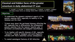
Classical and hidden faces of the greater omentum in daily abdominal CT scan. F. JAUSSET, B. COULIER, H. ROPION-MICHAUX, A. FAIRISE, V.LAURENT, D.REGENT CHU Nancy-Brabois Service de radiologie Adultes 54511 Vandoeuvre les Nancy Cedex . Objectives.

E N D
Classical and hidden faces of the greater omentum in daily abdominal CT scan F. JAUSSET, B. COULIER, H. ROPION-MICHAUX, A. FAIRISE, V.LAURENT, D.REGENT CHU Nancy-Brabois Service de radiologie Adultes 54511 Vandoeuvre les Nancy Cedex Objectives • To understand the dynamics anatomical aspects of the greater omentum (GO), especially its mobility in the abdomen and the chest • Tolearn more about the peritoneal diseases affecting the GO, especially how to differentiate the inflammatory greater omentum from the tumoral “omental cake” in peritoneal carcinomatosis and malignant mesothelioma • To be familiar with specific diseases of GO, especially ''parasitic'' benign or malignant and avoid diagnostic mistakes with there sometimes serious consequences. • - To remind semiological features of GO infarction.
1. Static and dynamic anatomy of the greater omentum GO and its arterial vessels :omental arteries and arcade of Barkow and Haller The greater omentum (GO) is, in our anatomical perception, arranged as an apron between the deep surface of the anterior abdominal wall and the mass of intestinal loops and their mesentery, hung on the transverse colon (red arrow) Embryologically, it is formed by the juxtaposition of primitive peritoneal layers coming from the stomach and the transverse colon. Thus, it is in continuity with the gastrocolic ligament and contribute by its upper edge to the constitution of the lower wall of the lesser sac.
COULIER B.64-row MDCT review of anatomic features and variations of the normal greater omentum Surg Radiol Anat 2009;31:489-500 B Coulier et al studied the length and the position of the right and left parts of the omental apron, based on the identification of omental vessels on "high resolution" CT and especially the veins that descend vertically into the GO from the greater curvature of the stomach.A measure of the maximum thickness of the subcutaneous fat was performed in each patient in front of the rectus abdominalis muscle at the level of the umbilicus A thorough statistical analysis of data collected in “normal” patients (no clinical history changing peritoneal images) and of image quality was performed.3D volumetric representations were made on selected cases to illustrate pedagogical aspects example of GO expanded, symmetrical, with excellent visibility of the vascular arcade of Haller and Barkow (arrow)
Example of short and thick GO COULIER B.64-row MDCT review of anatomic features and variations of the normal greater omentum Surg Radiol Anat 2009;31:489-500 Example of GO in the subphrenic and prehepaticspace example of GO preferentially developed in the right side of the abdomen
Main results of Coulier's study: COULIER B.64-row MDCT review of anatomic features and variations of the normal greater omentum Surg Radiol Anat 2009;31:489-500 -GO was identified and measured in all patients -in women, the left hemi-omentum was longer than the right one, and longer than left hemi-omentum of men -regarding males, there was no difference between the length of right and left hemi-GO, and no difference between men and women’s right hemi-GO -GO length was significantly higher in women than in men -the GO thickness in men was related to the subcutaneous thickness. Regarding the women, there was a predominance of the subcutaneous fat compared to the peritoneal fat -subphrenic GO is mainly encountered in men (32% for men vs 2% for women). This “curtain” of fat could explain the difficulties of the liver ultrasound in men GO completely in the leftside of the abdomen Subphrenic and prehepatic GO
Where is the GO The edematous GO isdrawninto an indirect inguinal hernia
Where is the GO GO ascendedinto a Morgagni’shernia.
Where is the GO The GO is in the supramesocolic space, « floating » in the ascitic fluid
2. Physiological and pathophysiological roles of the greater omentum [2] Roles of the GO as a part of the peritoneum : -support and protection -peritoneal fluid secretion -exchanges of water and ions with blood; colloids and particles with lymphatic vessels. Secretion and excretion concern protein fractions of the plasma (especially fibrin and mucin) and cells. Specific properties : -increase in volume (especially if inflammation) -regeneration (discussed) -mobility through the action of the diaphragm, the abdominal wall, bowel or changes in intraperitoneal pressure -adherence capacity, quick and accurate, a true chemotaxis, allowing GO to turn up to plug a perforated ulcer, an acute cholecystitis , or an anastomotic leak -defense by secretion of proteolytic enzymes -cellular defense by warning an adaptative vascularization -defense by antibody’s synthesis Acute cholecystis with GO’s plastron in the subhepatic space
3.Imaging analysis: inflammatory vs tumoral greater omentum -’’omental cake’’ is frequently used in radiological reports, but the definition should be clarified Tumoral ‘’omental cake’’: -density of tissue -retractile (fibrous) -progressive enhancement -with peritoneal nodules on the parietal layer (pelvis, paracolic gutter, subhepatic, right side of the diaphragm), the mesentery, easier to see in presence of ascitic fluid. -In absence of fluid, PET CT might be useful to detect small nodules
Inflammatory GO should not be called « omental cake » ‘’inflammatory’’ greater omentum: -large and diffuse thickening -heterogenous enhancement related to fatty areas with variable inflammatory changes (edema, granulomatous infiltration) -regular inflammatory thickening of the parietal peritoneum Tuberculous peritonitis diagnosis
4. « Parasitic » involvement of the greater omentum -pathologies that have migrated from another site (bowel, pelvis…) -different origins: .parasitic dermoïd cyst of the GO .parasitic GIST of the GO .parasitic leiomyoma of the GO .parasitic splenosis of the GO .carcinomatosis nodules could fall into this category of diseases that are fed by the GO!
58 years old woman with atypical abdominal pain diagnosis X-Ray, US and CT show calcified lesions with fat component (blue arrows) disseminated in the peritoneal cavity ; Ovarian dermoid cysts in the pelvis (green arrows) Parasitic dermoïd cyst of the GO from ovarian origin
71 years old man with palpable abdominal mass diagnosis Unifocal GIST of the GO, which origin can be gastric or intestinal
GIST of the GO are not exceptional, Miettinen and al described 95 cases proved by surgery in 2009. 21 were not attached to the gut. In this group, unifocal lesions were pathologically identical to the gastric GIST, sharing the same prognosis (a little better than intestinal lesions). In contrast, multiple lesions were identical to the intestinal GIST (mixed with epithelioïd component and Cajal cells) with the same poor prognosis. Unifocal omental involvements are probably gastric GIST migrated, while multifocal lesions of the GO, associated with intestinal GIST, are more probably metastases . Determination of the origin of a unifocal GO’s GIST (and its prognosis) requires the recognition of a macroscopic continuity with the gut (imaging, surgery or pathology)
43 years old woman. No surgical history. 4 children. Splenosis post-splenectomy for spleen traumatism 20 years before; intense and homogenous enhancement of splenosis nodules is characteristic; absence of symptom and gynecological history exclude endometriosis diagnosis
36 years old woman with abdominal pain diagnosis Small hypervascular nodules of the GO and the mesentery, first reflex : looking at the splenic space… When the spleen is missing, the diagnosis of splenosis is highly likely! ! !
5. ischemic, inflammatory and infectious lesions of the greater omentum 5.1 ischemic lesion of the GO Right hemi-GO infarction diagnosis
Laparoscopic aspect: partial torsion of the GO, with ischemic necrosis (blue arrow); important inflammatory response of the parietal peritoneum (yellow arrow) Infarction of the right GO: note the important inflammatory thickening of the parietal peritoneum (green arrows) Inflammatory response also involves the properitoneal fat and the abdominal wall (blue arrow). diagnosis
42 y.o woman. Abdominal pain after bariatric surgery diagnosis Appendagitis of the GO; cystic changes
diagnosis Ischemic epiploic hernia: strangulation in a rectus abdominalis muscles diastasis Note the important inflammatory response of the parietal peritoneum into the hernia.
5.2 Inflammatory lesions of the GO diagnosis Dermoïd peritonitis: 2 mature ovarian teratomas (blue arrows) including one ruptured causing acute chemical inflammatory peritonitis 39 years old woman. Acute abdominal pain 2 days before
49 years old woman. Acute abdominal pain and fever. Increasing of WBC diagnosis Gynecologic peritonitis (acute salpingitis); inflammatory edematous hypertrophy of the GO. The length of the GO in women explains its frequent involvement in gynecologic infection.
6. Primitive tumors of the greater omentum -all mesenchymal tumor types have been reported: malignant fibrous histiocytoma, liposarcoma, leiomyosarcoma, hemangiopericytoma / solitary fibrous tumor. -primitive mesothelioma of the GO can be a single mass; tissular or cystic but mostly disseminated. -primitive location of the hematologic malignancies: non Hodgkin lymphoma, granulocytic sarcoma can also be a single mass but mostly are disseminated diffusion diagnosis 52 years old man, abdominal mass
PEComa of the GO -diagnosed by immunohistochemistry : sarcomatous lesion containing epithelioid cells with smooth muscle (smooth muscle actin) and melanocytic biomarker (HBM 45, melan A) -PEComa is a proliferation of perivascular epithelial cells (periepitheloid cells=PEC) encountered in: .sarcoma with perivascular epithelioid cells .clear cell myomelanocytic tumor .clear cell « sugar » tumor .renal angiomyolipoma with clear cells and usual renal or liver angiomyolipoma .lymphangioleiomyomatosis Smooth muscle actine HMB45
diagnosis 20 years old woman with RLQ pain and fever Myofibroblastic tumor of the GO (inflammatory pseudotumor) -atthisage, differentialdiagnosisincludes: .desmoplastic small round cells tumor (PNET primary neuroectodermal tumor) .hemangiopericytoma/solitaryfibrous tumor .Castelmandisease …non Hodgkin and Hodgkin lymphoma are not as hypervascular; … granulocyticsarcoma in hematologicmalignancies (acute myeloidleukemia M4 and M5).
7. Secondary tumoral involvement of the GO -the '’’omental cake’’ is easy to identify when ascitic fluid is present; coexistence with regular or irregular thickening of the parietal peritoneum and mesenteric retraction. -Without ascitic fluid, it is more challenging to detect peritoneal nodules. Indeed, PET CT daily reminds us that we have to carefully look at all peritoneal nodules Cephalic pancreatic adenocarcinoma with carcinomatosis -in this case no relation exists between the volume of the ascite and the size of the peritoneal nodules (blocking of the cisterna chyli by the posterior extension of pancreatic cancer). -nodules of the GO are in the supramesocolic space ! ! !
Gastric linitis; peritoneal and hepatic pedicle dissemination ;main bile duct endoprosthesis -minor involvement of the greater omentum related to the important mesenteric retraction -no macroscopic peritoneal nodules on the parietal peritoneum, despite the abundant ascitic fluid
27 years old woman, abdominal pain diagnosis -sometimes the appearance of omental lesions may be characteristic of an origin -psammomatous calcifications of the GO (yellow arrows), and of the prehepatic parietal peritoneum (green arrow) are typical of a serous papillary ovarian carcinoma -primitive bilateral lesion shows "sand-like"calcifications Courtesy Dr Ranchoup Clinique du Mail Grenoble
7. Miscellaneous lesions …..of the GO 59 yo man, loss of weight, chronic diarrhea, cardiac insuffisiency diagnosis Primitive amyloidosis Calcified nodules in the GO suggest a chronic disease: tuberculosis or amyloidosis
87 years old woman with acute abdominal pain and obstruction diagnosis -defect of enhancement of the small bowel’s wall caused by arterial vessel’s strangulation. -convergence of mesenteric folds of the distended loops and their vessels to the anterior part of the RLQ (“C” disposition of the loops) -caecum pushed backwards and inwards, confirming the anterior location of the small bowel loops - GO is not visible between the loops and the abdominal wall Transomental hernia Strangulated transomental hernia: CT findings E. DELABROUSSE, M. COUVREUR, O. SAGUET, B. HEYD, S. BRUNELLE, B. KASTLER Abdominal Imaging ; 2001,26:89-88 Courtesy of Pr Delabrousse C HU Besançon - France
76 years old woman with acute abdominal pain and obstruction diagnosis • -radial disposition of the distended loops in the pelvis; no enhancement of the bowel wall (arterial vessels strangulation). • - convergence of mesenteric folds of the distended loops and their vessels to the anterior part of the abdomen • The GO is not visible between the loops and the abdominal wall • -hole at the lower part of the GO Transomental hernia CT of internal hernias TAKEYAMA N, GOKAN T, OHGIYA Y, SATOH S, I HASHIZUME, MD, HATAYA K., KUSHIRO H., NAKANISHI M., KUSANO M., MUNECHIKA H., RadioGraphics ;2005,25:997-1015
7. Conclusion -the GO is easy to identify when it’s enlarged by inflammatory, infectious, ischemic or tumoral disease -we have to keep in mind its mobility that can cause misdiagnosis -abnormalities of the greater omentum should always be confronted with clinical, epidemiological and pathological data to suggest a panel of realistic diagnostic hypothesis
Bibliography 1.Hollender L, Bur F, Pigache P. chirurgie du grand epiploon. In:EMC Techniques chirurgicales -Appareil digestif, 2002. 2 Coulier B. 64-row MDCT review of anatomic features and variations of the normal greater omentum. Surg Radiol Anat 2009; 31:489-500. 3Yoo E, Kim JH, KimMJ, et al. Greater and lesser omenta: normal anatomy and pathologic processes. Radiographics 2007; 27:707-720. 4.Jin H, Min PQ, Yang ZG, Song B, Wu B. A study of multi-detector row CT scan on greater omentum in 50 individuals: correlating with anatomical basis and clinical application. Surg Radiol Anat 2008; 30:69-75. 5Liebermann-Meffert D. The greater omentum. Anatomy, embryology, and surgical applications. Surg Clin North Am 2000; 80:275-293, xii. 6.Coulier B, Pringot J. [Pictorial essay. Infarction of the greater omentum: can US and CT findings help to avoid surgery?]. JBR-BTR 2002; 85:193-199. 7.van Breda Vriesman AC, Puylaert JB. Epiploic appendagitis and omental infarction: pitfalls and look-alikes. Abdom Imaging 2002; 27:20-28. 8.Fasih N, Prasad Shanbhogue AK, Macdonald DB, et al. Leiomyomas beyond the uterus: unusual locations, rare manifestations. Radiographics 2008; 28:1931-1948. 9.Cohen DT, Oliva E, Hahn PF, Fuller AF, Jr., Lee SI. Uterine smooth-muscle tumors with unusual growth patterns: imaging with pathologic correlation. AJR Am J Roentgenol 2007; 188:246-255. 10.Takeda A, Mori M, Sakai K, Mitsui T, Nakamura H. Parasitic peritoneal leiomyomatosis diagnosed 6 years after laparoscopic myomectomy with electric tissue morcellation: report of a case and review of the literature. J Minim Invasive Gynecol 2007; 14:770-775. 11.Franzini C, Alessandri L, Piscioli I, et al. Extra-gastrointestinal stromal tumor of the greater omentum: report of a case and review of the literature. World J Surg Oncol 2008; 6:25. 12.Nakagawa M, Akasaka Y, Kanai T, et al. Extragastrointestinal stromal tumor of the greater omentum: case report and review of the literature. Hepatogastroenterology 2003; 50:691-695. 13.Miettinen M, Sobin LH, Lasota J. Gastrointestinal stromal tumors presenting as omental masses--a clinicopathologic analysis of 95 cases. Am J Surg Pathol 2009; 33:1267-1275. 14.Shin MK, Lee OJ, Ha CY, Min HJ, Kim TH. Malignant mesothelioma of the greater omentum mimicking omental infarction: a case report. World J Gastroenterol 2009; 15:4856-4859. 15 Ben Achour J, Bouasker I, Bedoui R, et al. [Myofibroblastic tumor of the great omentum]. Tunis Med 2008; 86:932-935 16.Delabrousse E, Couvreur M, Saguet O, Heyd B, Brunelle S, Kastler B. Strangulated transomental hernia: CT findings. Abdom Imaging 2001; 26:86-88. 17 Coulier B, Elst BV, Pierard F. Closed loop small bowel occlusion through a congenital defect of the greater omentum. JBR-BTR; 93:106