Download
1 / 105
1.08k likes | 1.24k Views
Bones Of The Leg Popliteal Fossa The Knee. Dr. Fadel Naim Orthopedic Surgeon IUG. PATELLA. The largest sesamoid bone Triangular Its apex lies inferiorly The posterior surface articulates with the condyles of the femur. PATELLA.

E N D
Bones Of The Leg Popliteal FossaThe Knee Dr. Fadel Naim Orthopedic Surgeon IUG
PATELLA • The largest sesamoid bone • Triangular • Its apex lies inferiorly • The posterior surface articulates with the condyles of the femur
PATELLA • Connected to the tuberosity of the tibia by the ligamentum patellae. • It is prevented from being displaced laterally during the action of the quadriceps muscle by: • The lower horizontal fibers of vastus medialis • The large size of the lateral condyle of the femur
Patella • Supported by muscle bone and ligamentous structures • Muscle- through quad tendon • Medially- vastus medialis • Laterally- vastus lateralis • Superiorly- rectus femoris and vastus intermedius • Bone- • Trochlear groove • Ligamentous- • Patellar ligament • Patellar retinacula • Lateral • Medial
Function • Increased efficiency of quadriceps • Changes line of pull • Protection of anterior knee joint
Functions of Patello-femoral Joint with patella without patella (1) increases angle of pull of quads on tibia, improves the ratio of motive:resistive torque by 50% (2) centralizes divergent tension of quads into a single line of action (3) some protection of anterior aspect of knee
Patella • Exposed position in front of the knee joint and can easily be palpated through the skin. • It is separated from the skin by an important subcutaneous bursa
Tibia • The large weight-bearing medial bone of the leg • It articulates with: • The condyles of the femur • The head of the fibula • The talus • The distal end of the fibula • It has an expanded upper end, a smaller lower end, and a shaft.
Tibia • At the upper end: • The lateral and medial condyles (sometimes called lateral and medial tibial plateaus), • Articulate with the lateral and medial condyles of the femur • Anterior and posterior intercondylar areas separate the upper articular surfaces of the tibial condyles • Intercondylar eminence lies between these areas
The lateral condyle possesses on its lateral aspect a small circular articular facet for the head of the fibula. • The medial condyle has on its posterior aspect the insertion of the semimembranosus muscle
Proximal Tibia Anatomy • Lateral tibial Plateau: convex, smaller than medial plateau • Lateral intercondylar eminence • Medial intercondylar eminence • ------------------------------------- • Medial tibial plateau: concave, larger than lateral plateau • Tibial tubercle: insertion of patellar tendon • Tibial shaft • Fibular shaft • Fibular head: Styloid process of fibular head is the incertions of the lateral collateral ligament. • Gerdy's tubercle: insertion site of iliotibial band
The Shaft Of The Tibia • Triangular in cross section • Three borders and three surfaces • Anterior, medial borders and the medial surface are subcutaneous. • The anterior border is prominent and forms the the shin. • At the junction of the anterior border with the upper end of the tibia is the tuberosity, • Receives attachment of the ligamentum patellae. • The anterior border becomes rounded below, where it becomes continuous with the medial malleolus
The Shaft Of The Tibia • The lateral or interosseous border gives attachment to the interosseous membrane • The posterior surface of the shaft shows an oblique line, the soleal line for the attachment of the soleus muscle
The lower end of the tibia is slightly expanded and on its inferior aspect shows a saddle-shaped articular surface for the talus. • The lower end is prolonged downward medially to form the medial malleolus. • The lateral surface of the medial malleolus articulates with the talus. • The lower end of the tibia shows a wide, rough depression on its lateral surface for articulation with the fibula.
FIBULA • The slender lateral bone of the leg • No part in the articulation at the knee joint • Below it forms the lateral malleolus of the ankle joint. • No part in the transmission of body weight • Provides attachment for muscles. • An expanded upper end, a shaft, and a lower end.
The upper end, or head • A styloid process. • Articular surface for articulation with the lateral condyle of the tibia • The shaft of the fibula • Long and slender. • Four borders and four surfaces • The medial or interosseous border gives attachment to the interosseous membrane.
The lower end of the fibula • Forms the triangular lateral malleolus, which is subcutaneous. • On the medial surface of the lateral malleolus is a triangular articular facet for articulation with the lateral aspect of the talus. • Below and behind the articular facet is a depression called the malleolar fossa.
PATELLAR DISLOCATIONS • Congenital recurrent dislocations • caused by underdevelopment of the lateral femoral condyle. • Traumatic dislocation of the patella • results from direct trauma to the quadriceps attachments of the patella (especially the vastus medialis), with or without fracture of the patella.
PATELLAR FRACTURES • A result of direct violence • Broken into several small fragments • Because the bone lies within the quadriceps femoris tendon, little separation of the fragments takes place. • The close relationship of the patella to the overlying skin may result in the fracture being open. • A result of indirect violence • Caused by the sudden contraction of the quadriceps • Snapping the patella across the front of the femoral condyles. • The knee is in the semiflexed position • The fracture line is transverse • Separation of the fragments usually occurs.
FRACTURES OF THE TIBIA • Fractures of the tibia and fibula are common. • If only one bone is fractured, the other acts as a splint and displacement is minimal. • Fractures of the shaft of the tibia are often open because the entire length of the medial surface is covered only by skin and superficial fascia.
FRACTURES OF THE TIBIA • Fractures of the distal third of the shaft of the tibia are prone to delayed union or nonunion. • This can be because the nutrient artery is torn at the fracture line, with a consequent reduction in blood flow to the distal fragment; • The splint like action of the intact fibula prevents the proximal and distal fragments from coming into apposition.
Fractures of the proximal end of the tibia (tibial plateau) • Common in the middle aged and elderly • Usually result from direct violence to the lateral side of the knee joint • The tibial condyle may show: • A split fracture • Be broken up • The fracture line may pass between both condyles in the region of the intercondylar eminence. • As a result of forced abduction of the knee joint, the medial collateral ligament can also be torn or ruptured.
INTRAOSSEOUS INFUSION OF THE TIBIA IN THE INFANT • For the infusion of fluids and blood when it has been found impossible to obtain an intravenous line. • The bone marrow needle is directed at right angles through the skin, superficial fascia, deep fascia, and tibial periosteum and the cortex of the tibia. • Once the needle tip reaches the medulla and bone marrow, the operator senses a feeling of "give." • The position of the needle in the marrow can be confirmed by aspiration. • The transfusion may then commence.
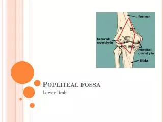
Popliteal Fossa • a diamond-shaped intermuscular space situated at the back of the knee • most prominent when the knee joint is flexed.
Popliteal Fossa • It contains: • The popliteal vessels • The small saphenous vein • The common peroneal nerve • Tibial nerve • The posterior cutaneous nerve of the thigh • The genicular branch of the obturator nerve • Connective tissue • Lymph nodes
Popliteus • A thin, triangular muscle • Forms the inferior part of the floor of the popliteal fossa • The apex of its fleshy belly emerges from the joint capsule of the knee joint. • Origin: • posterior lateral condyle of femur • Insertion: • upper posterior medial surface of tibia • Action • flex knee, internally rotate knee
POPLITEAL ARTERY • deeply placed and enters the popliteal fossa through the opening in the adductor magnus • ends at the level of the lower border of the popliteus muscle by dividing into anterior and posterior tibial arteries.
POPLITEAL ARTERY • Relations • Anteriorly: • The popliteal surface of the femur • the knee joint • the popliteus muscle • Posteriorly: • The popliteal vein • the tibial nerve • Fascia • skin • Branches • muscular branches • articular branches to the knee.
POPLITEAL ANEURYSM • The pulsations of the wall of the femoral artery against the tendon of adductor magnus at the opening of the adductor magnus is thought to contribute to the cause of popliteal aneurysms.
SEMIMEMBRANOSUS BURSA SWELLING • The most common swelling found in the popliteal space. • It is made tense by extending the knee joint and becomes flaccid when the joint is flexed. • A baker's cyst • Centrally located • Arises as a pathologic (osteoarthritis) diverticulum of the synovial membrane through a hole in the back of the capsule of the knee joint.
POPLITEAL VEIN • Formed by the junction of the venae comitantes of the anterior and posterior tibial arteries at the lower border of the popliteus muscle • It begins on the medial side of the popliteal artery. • As it ascends through the fossa, it crosses behind the popliteal artery so that it comes to lie on its lateral side • It passes through the opening in the adductor magnus to become the femoral vein. • Tributaries • Veins that correspond to branches given off by the popliteal artery. • Small saphenous vein, which perforates the deep fascia and passes between the two heads of the gastrocnemius muscle to end in the popliteal vein. • Popliteal fossa • Semitendinosus • Biceps femori • Semimembranosus • Sciatic nerve • Popliteal vein • Popliteal artery
ARTERIAL ANASTOMOSIS AROUND THE KNEE JOINTGenicular anastomosis • To compensate for the narrowing of the popliteal artery, which occurs during extreme flexion of the knee • around the knee joint is a profuse anastomosis of small branches of: • The femoral artery • Muscular and articular branches of the popliteal artery • Branches of the anterior and posterior tibial arteries .
TIBIAL NERVE • The larger terminal branch of the sciatic nerve • The tibial nerve arises in the lower third of the thigh. • It runs downward through the popliteal fossa, lying first on the lateral side of the popliteal artery, then posterior to it, and finally medial to it • The popliteal vein lies between the nerve and the artery throughout its course. • The nerve enters the posterior compartment of the leg by passing beneath the soleus muscle.
COMMON PERONEAL NERVE • The smaller terminal branch of the sciatic nerve • arises in the lower third of the thigh • It runs downward through the popliteal fossa closely following the medial border of the biceps muscle • It leaves the fossa by crossing superficially the lateral head of the gastrocnemius muscle. • It then passes behind the head of the fibula, winds laterally around the neck of the bone • it is subcutaneous and can easily be rolled against the bone
Knee Dr. Fadel Naim Orthopedic Surgeon Faculty of medicine IUG