Download

1 / 19
190 likes | 342 Views
PhD Course. TOPICS IN (NANO) BIOTECHNOLOGY Lecture V. 29th April, 2003. DNA Microarrays - Mechanics -. Orderly, high-density arrangements of nucleic-acid spots (probes) immobilized on a suitable substrate (e.g., glass). Hundreds of identical arrays are often “printed” together.

E N D
PhD Course TOPICS IN (NANO) BIOTECHNOLOGY Lecture V 29th April, 2003
DNA Microarrays - Mechanics - • Orderly, high-density arrangements of nucleic-acid spots (probes) immobilized on a suitable substrate (e.g., glass). • Hundreds of identical arrays are often “printed” together. • Probes may be of two different types: • 1) gene (cDNA) fragments; • 2) oligonucleotides. • Probe deposition is computer aided with up to 10,000 cDNA or 250,000 oligonucleotide spots/cm2 being routinely used today. • The “target” mRNA is reverse-transcribed into cDNA and either radiolabeled or labeled with fluorescent molecules for detection. • Fluorescence is the method of choice. • Fluorescence analysis requires the use of two target samples. • Once labeled, the cDNA pools are hybridized to the DNA array and the signals are detected by a “reader”. • Data is analyzed with sophisticated bioinformatics software to resolve, quantitate and group the signals in a meaningful way.
General Overview of cDNA Microarray Process Microarray Preparation cDNA Probe Hybridization Confocal Microscope Computer Analysis
2D Graph showing genes which were highly regulated relative to control in the 50 and 2.5 mg/kg drug treatments. Drug 50 mg/kg 25 mg/kg 2.5 mg/kg 0.25 mg/kg T-ceIl specific protein Putative purine nucleotide binding protein Mtrp1 beta variant (Mtrp1) Mus musculus nimA-reIated kinase 2 (Nek2) Mouse mRNA for interleukin-6 Mouse testosterone 16-alpha-hydroxylase Mouse testosterone 16-alpha-hydroxyIase (C-16-alpha) ATP Citrate Lyase Cyp4a-10 SODIUM/BILE ACID COTRANSPORTER LOW MOLECULAR WEIGHT GROWTH HORMONE RECEPTOR/BI “FRUCTOSE-1, 6-BISPHOSPHATASE” Mouse histidase locus histidine ammonia-lyase Group 1 major urinary protein Mus musculus reelin Cytochrome P450 monooxygenase (CYP2J5) M.musculus spot14 gene.
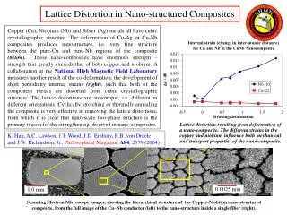
Graph showing expression changes for genes that have been associated with hepatotoxicity. Genes that are colored in red were upregulated relative to control, genes that are colored green were downregulated relative to control. (B). Table showing the expression changes of genes normally regulated during an immune response. Drug 0.25 mg/kg 2.5 mg/kg 50 mg/kg 25 mg/kg CYP4B1 M. musculus Cyp4 a-10 mRNA Cytochrome P450 (Cyp1b1) Cytochrome P450 Cyp7b1 Cytochrome P450 monooxygenase (CYP2J5) Cytochrome P450 monooxygenase (CYP2J6) M.musculus CYP1A1 gene CYP4A14 Glutathione S-transferase Glutathione Peroxidase Glutathione Synthetase UDP GIucuronosyltransferase Acetyl Transferease Proliferating cell nuclear antigen CycIin B1 Cyclin B2 Cyclin A CycIin E
100000 10000 Induced by Oligonucleotide Treatment Drug 1000 Inhibited by Oligonucleotide Treatment 100 RNA Expression 10 10 100 1000 10000 100000 RNA Expression Drug DNA Array Analysis of Drug Treated Cells
DNA Microarrays- Limitations - • Need for standardization of the technology to enable comparisons of data between labs. • Large amount of high quality sample RNA is generally required (50-200 g). Sensitivity limitations. • Reproducibility of chip quality, wet methodology. • Expensive and labor intensive. • mRNA levels may not parallel protein levels, protein activity.
Proteomics I • Definitions • Proteome: The total protein complement of a genome (1995). • Proteomics: A scientific discipline devoted to defining and characterizing the proteomes of specific organisms. • Proteomics is inherently more complex than DNA-based (e.g., arrays) technologies. • Larger alphabet (4 vs 20) • RNA splicing and editing produces different proteins from a single gene. • Proteins are post-translationally modified in many different ways (e.g., spliced, phosphorylation). • Methods for separating and detecting proteins is more complex. • Proteomics is still largely centered upon two-dimensional (2-D) gel electrophoresis. • Been in existence >25 years • The only method currently available which is capable of simultaneously separating and resolving thousands of proteins.
Sequence motifs for some of the common co- and post-translational modifications. Amino acids in italicsare those that will be modified in each case
Proteomics II • The concept of 2-D gels • Proteins are separated first based on charge (1st dimension): • Isoelectric Focusing (IEF). • Proteins are separated in a pH gradient until they reach a point in which their net change = 0 (isoelectric points or pI). • Following IEF proteins are next separated based on molecular mass (2nd dimension). • Presence of sodium dodecyl sulphate (SDS) overrides the intrinsic change of a protein. • Recent advances in 2-D gel electrophoresis, protein microanalysis and bioinformatics have made the large-scale, systematic analysis of proteins possible. • Immobilized pH gradients (IPGs) • Replaces soluble ampholytes to maintain charge gradient. • Permits much larger amounts of protein to be analyzed.
Proteomics III • Commercially-available precast gels of sufficient quality permits reproducibility. • Major advances in Mass Spectrophotometry. • Identification of amino acid sequence and post-translational modifications based on mass. • Highly sensitive. • Bioinformatics permits the large-scale analysis of data over a reasonable time-frame. • Proteomics databases are widely available today on the web to support proteomics research.
2-Dimensional Gel Electrophoresis pH 3.5 pH 10.0 96 kDa 14 kDa
Protein Analysis Based on 2-D Gel Technology A B Electrotransfer ontomembrane Vector analysis Excise protein spot from the gel In situ proteinase digest (peptides passively eluted into supernatant) Acid hydrolyses (monosaccharide, phosphoamino & amino acid composition) Endoproteinase digest of protein Enzymic release of modifications High sensitivity Edman sequencing MS Analysis m/z 366 (HexNAc-Hex+) m/z 204 (HexNAc+) m/z 292 (NeuAc+) m/z 79 (PO3-) fragment ions EdmanSequence
Proteomics- Limitations - • Expensive and labor intensive. • Non-specific protein contamination due to ultra-sensitivity of new detection methods. • Protein levels may not parallel protein activity. • Standardization of technology to enable comparative studies between different investigators still in progress.
Target Validation Approaches TargetValidation Drug Identification Clinical Development Gene Protein Correlative Approaches Causative Approaches • Comparative Genomics • Transcriptional Profiling • Proteomics • Overexpression Systems • Gene Knockouts • Small Molecules • Antibody Approaches • Antisense • Interference RNA