Download

1 / 32
340 likes | 1.01k Views
Methods to Detect Red Cell Membrane Disorders. Practical Clinical Hematology. OSMOTIC FRAGILITY TEST. 10. Definition. The osmotic fragility test is a measure of the ability of the red cells to take up fluid without Lysing.

E N D
Methods to Detect Red Cell Membrane Disorders Practical Clinical Hematology OSMOTIC FRAGILITY TEST 10
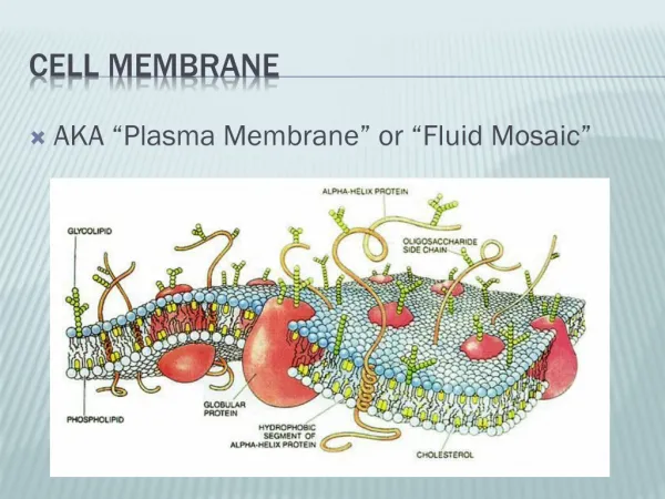
Definition • The osmotic fragility test is a measure of the ability of the red cells to take up fluid without Lysing. • The osmotic fragility test is a measure of the resistance of erythrocytes to hemolysis by osmotic stress (exposed to a series of increasingly dilute saline solutions). • The sooner hemolysis occurs, the greater the osmotic fragility of the cells.
Factors affecting on the osmotic fragility • The primary factor affecting the osmotic fragility test is the shape of the red cell, which, in turn, depends on the • Volume • Surface area • Functional state of the red blood cell membrane.
Increased surface- to – volume ratios • It is more resistant to hemolysis and has decreased fragility • The larger the amount of red cell membrane (surface area) in relation to the size of the cell, the more fluid the cell is capable of absorbing before rupturing . as • Reticulocytes • Iron-deficiency anemia • Thalassaemia • Sickle cell anemia • and occurs following splenectomy, in liver disease, polycythemia vera, and conditions in which target cells are present.
The target cell has the largest surface area (amount of membrane) for its size and therefore shows decreased fragility. • As the red cell takes in fluid it becomes more round (spherocytic). It therefore follows that the spherocyte has the smallest surface area for its volume, ruptures the most quickly, and has increased fragility.
Decreased surface-to – Volume ratios • Increased osmotic fragility (decreased resistance) is found in • Hemolytic anemias • Hereditary Spherocytosis • And whenever Spherocytes are found. • The older red cells are also more fragile.
Specimen • Heparinized venous blood , or, 15 to 20 mL of defibrinated whole blood. • The test should be set up within 2 hours of collection, or within 6 hours if the blood is refrigerated. Reagent • Stock Solution of buffered sodium chloride NaCl 90 g • Na2HPO4 13.65 g or Na2HPO4.H2O 17.115 g • NaH2P04.2H2O 2.34g • Water to 1 L • From (the stock solution, prepare first a 10 g/L solution by dilution with water. Dilutions equivalent to 9.0, 7.5, 6.5, 6.0, 5.5, 5.0, 4.0, 3.5, 3.0, 2.0, 1.0 g/L are then prepared.
Principle • In the osmotic fragility test, whole blood is added to varying concentrations of buffered sodium chloride solution and allowed to incubate at room temperature. • The amount of hemolysis in each saline concentration is then determined by reading the supernatants on a spectrophotometer.
Procedure • Prepare dilutions of buffered sodium chloride and place in the appropriately labeled test tube (See Table). • Mix the preceding dilutions well, using Para film to cover each test tube while mixing. • Transfer 5 mL of each dilution to a second set of test tubes labeled # 1 through #14. • Add 0.05 mL of the patient's Heparinized blood to each of the 14 test tubes. Repeat, adding the normal control blood to the set of 14 control test tubes. • Mix each test tube immediately by gentle inversion.
Allow the test tubes to stand at room temperature for 30 minutes. • Remix the test tubes gently and centrifuge at 1200 to 1500 g for 5 minutes. • Carefully transfer the supernatants to cuvettes and read on a spectrophotometer at a wavelength of 540 nm. Set the optical density at 0, using the supernatant in test tube #1, which represents the blank, or 0% hemolysis. Test tube #14 represents 100% hemolysis. • Calculate the percent hemolysis for each supernatant as follows:
The results of the test may then be graphed, with the percent hemolysis plotted on the ordinate (vertical axis) and the sodium chloride concentration on the abscissa (horizontal axis) as shown in Figure (shows normal range). Osmotic fragility can be described in terms of the saline concentration at which lysis begins • initial lysis or minimum resistance normally 4.5 - 5.0 g/L) • Complete lysis or maximum resistance, normally 3.0 - 3.3 g/L). • It is essentially useful to record the concentration of saline causing 50 % lysis, i.e. the Median Corpuscular Fragility (MCF), normally 4,0 - 4.45 g/L. and to inspect the entire curve.
Discussion • Presence of hemolytic organisms in the sample. • Severe anemia or other conditions with fewer RBCs available for testing . • Recent blood transfusion. • Old sample. • If anticoagulated blood is used for test, use only heparin as the anticoagulant, in order to avoid adding more salts to the blood such as Oxalate, EDTA, or citrate.
Discussion • Three variables capable of markedly affecting the results, apart from the accuracy of saline concentrations: • The relative volumes of blood and saline. • The final pH of the blood in saline suspension (should be 7.4%) • The temperature at which the tests are carried out. • Completely fill the collection tube and invert it gently several times to mix the sample and anticoagulant thoroughly. • Handle the sample gently to prevent accidental hemolysis.
Practical Clinical Hematology G6PD 11
Gluose-6-phosphate dehydrogenase (G-6-PD) • G6PD deficiency is the most common human enzyme deficiency in the world; it affects an estimated 400 million people. • G6PD deficiency is also known as "favism," since G6PD deficient individuals are also sometimes allergic to fava beans. • G6PD deficiency is an allelic abnormality which is inherited in an X-linked recessive fashion.
The complications can arise; hemolytic anemia and prolonged neonatal jaundice are the two major pathologies associated with G6PD deficiency. Both of these conditions are directly related to the inability of specific cell types to regenerate reduced nicotinamide adenine dinucleotide phosphate (NADPH); this reaction is normally catalyzed by the G6PD enzyme. • In G6PD deficient individuals, anemia is usually caused by certain oxidative drugs, infections, or fava beans. When any one of these agents, or their metabolites, enters a G6PD deficient red blood cell, hemoglobin becomes denatured, thus destroying its function as the principal oxygen carrying molecule.
In addition to being susceptible to hemolytic anemia, G6PD deficient individuals are also predisposed to prolonged neonatal jaundice. This can be a potentially serious problem as it can cause severe neurological complications and even death.
Principle: • Glucose-6-phosphate dehydrogenase (G6PDH, D-glucose-6-phosphate) catalyzas the first step in the pentose phosphate shunt ,oxidising glucose-6-phosphate (G-6-P)to 6-phosphogluconate(6-PG) and reducing NADP to NADPH,which illustrate by the following equation:
NADP is reduced by G-6-PDH in the presence of G-6-P. The rate of formation of NADPH is directly proportional to the G-6-PDH activity and is measured spectrophotometrically as an increased in absorbance at 340nm. Prodution of asecond molar equivalant of NADPH by erythrocyte 6-phosphogluconate dehydrogenase (6-PGDH) according to the reaction :
Specimen collection and storage • Whole blood collected with EDTA, or acid citrate dextrose . • Red cell G-6-PDH is stable in whole blood for one week refrigrated (2-8ºc),but is unstable in red cell hemolysates. since activity is reported in term of number of red blood cell or gram hemoglobin, the red cell count or hemoglobin concentration should be determined prior to performing the G6PDH assay.
Procedure • The temperature of the reaction mixture should be maintained at 30ºc or some other constant temperature. • prepare reaction mixture: • Add 0.01ml blood directly to vial containing G-6-PDH assay solution and mix thoroughly to completely suspend erythrocytes, let stand at room temperature (18-25ºc) for 5-10min. • Add 2.0ml G-6-PDH substrate solution directly to vial and mix gently by inverting several times. • Transfer contents of vial to cuvette. • Place cuvette in constant temperature cuvette compartment or water bath and incubate forapproximately 5min to attain thermal equilibrium.
Read and record absorbance (A1) of test at 340 nm vs. water or potassium dichromatesolution. This is initial A .(if using a water bath or incubator ,return cuvette to it) • Exactly 5 min later, again read and record (A2), this is final A. • To determine G-6-PDH activity do the following calculation.
Calculation: • ΔA per min= A2-A1/5 • G-6-PDH activity is expressed as U/1012 erythrocyte (RBC)or as U/g hemoglobin (Hb). • G-6-PDH (U/1012 RBC) = ΔA per min X 3.01 X 10¹²X TCF / 0.01 X 6.22 X (N X 10*6) X 1000 • Where: 3.01 = total reaction volume (ml) 1012 = factor for expressing activity in 1012 cells 0.01 = sample volume (ml) 6.22 = millimolar absorptive of NADPH at 340 nm 1000 = conversion of red cell count from mm³ to ml TCF = temperature correction factor (1at 30ºc) N X 106 = red cell count (red cells/mm³) determined for each specimen
This equation reduced to: G-6-PDH ( U/1012 RBC)= ΔA/min X (48,390/N) X TCF • Where: N = red cell count divided by 106 TCF = temperature correction factor (1at 30ºc) • G-6-PDH(U/g Hb) = ΔA per min X 100 X 3.01 / ((0.01 X 6.22 X Hb(g/dl)) X TCF = ΔA per min X 4839 / Hb (g/dl) X TCF • Where: 100 = factor to convert activity to 100ml 3.01 = total reaction volume (ml) 0.01 = sample volume (ml) 6.22 = mill molar absorptive of NADPH at 340 nm Hb (g/dl) = hemoglobin concentration determined for each specimen TCF = temperature correction factor (1 at 30ºc) • Note: If anemia and/or leukocytosis is present:Use buffy coat free blood sample for assay (platelets and WBCs marked activity in this enzyme)
Normal range: • G-6-PDH (U/1012 RBC): (146-376) • G-6-PDH (U/g Hb): (4.6-13.5)
Qualitative method in G-6-DP determination • Principle Glucose -6-phosphate dehydrogenase, present in the red blood cell hemolysate, act on glucose -6-phosphate and reduces NADP to NADPH which, with the help of PMS, reduces blue colored 2,6-Dichlorophenol Indophenol into a colorless form. the rate of declorization is proportional to the enzyme activity. The reaction can be represented as:
Note: • Fresh blood sample should be use since refrigeration reduces the enzyme activity. • Heparin sample should not be use as interfere with enzyme reaction. • Avoid exposure of substrate vial to the light (it is photosensitive).
Procedure: • Step1: Preparation of red cell hemolysate: • Purified water : 2.5ml • Fresh blood : 0.05ml • Mix well and allow standing for 5min at R.T. • Step2: Assay of the enzyme: • Add 1ml of the hemolysate (step 1) to the vial of solution 1 and mix gently. • Add immediately about 1ml of reagent 3. • Seal the vial with aluminum foil and incubate in water bath at 37ºc. • Observe:the time taken for the color change from initial deep blue to reddish purple. Follow up to a max. Of 6 hours with 30 min intervals.
Results: • Normal : 30-60 min. • G-6-PD deficient (hemizygous males, homozygous female): 140 min-24hr • G-6-Pd carriers (heterozygous females): 90 min-several hours.