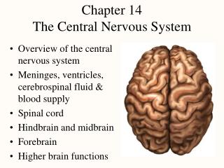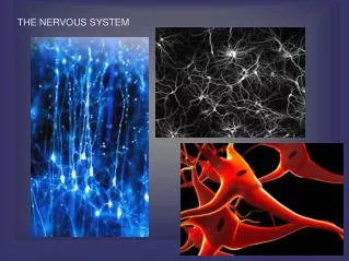
Nervous System: The Brain
630 likes | 842 Views
Nervous System: The Brain. Embryonic Development. Nervous system develops from ectoderm by 3rd week, neural plate becomes a groove with neural folds along each side by 4th week, neural folds join to form neural tube

Nervous System: The Brain
E N D
Presentation Transcript
Embryonic Development • Nervous system develops from ectoderm • by 3rd week, neural plate becomes a groove with neural folds along each side • by 4th week, neural folds join to form neural tube • lumen of the neural tube develops into central canal of spinal cord and ventricles of the brain • cells along the margin of the neural groove is called the neural crest • develop into sensory and sympathetic neurons and schwann cells • by 4th week, neural tube exhibits 3 anterior dilations
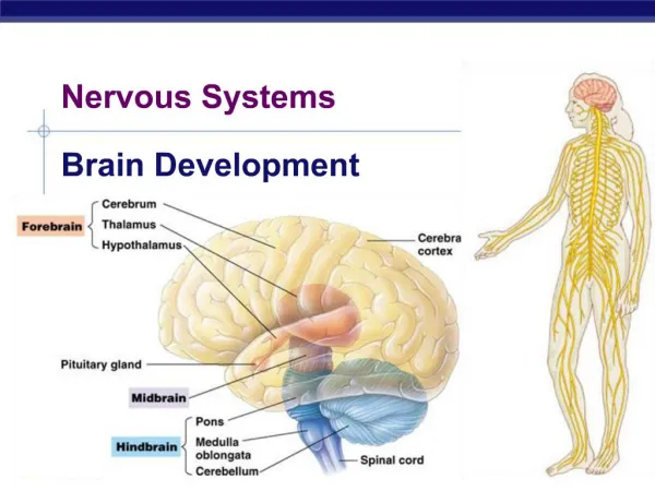
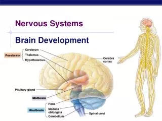
Embryonic Brain Development • 4th week • forebrain • midbrain • hindbrain • 5th week • telencephalon • diencephalon • mesencephalon • metencephalon • myelencephalon
FOREBRAIN Telecephalon: Cerebral Hemispheres, (cerebral cortex,basal nuclei, amydoloid nucleus) Diencephalon MIDBRAINmesencephalon HINDBRAIN(RHo mbencephalon) Metencephalon (Pons and cerebellum) Myenlencephalon (Medulla Oblongata) Gross Anatomy of the Brain
It is estimated that there are 100 billion (100,000,000,000) neurons in the human brain!
Human Brain Development • The Neuron Connection….. • At times during brain development, 250,000 neurons are added every minute!! At birth, almost all the neurons that the brain will ever have are present. However, the brain continues to grow for a few years after birth. By the age of 2 years old, the brain is about 80% of the adult size. • You may wonder, "How does the brain continue to grow, if the brain has most of the neurons it will get when you are born?". The answer is …
Answer… • Glial Cells. Glia continues to divide and multiply • The neurons in the brain also make many new connections after birth. The brain, thus, has PLASTICITY, or NEUROPLASTICITY…a hot topic in neurobiological research today.
Notice the Crista Galli marked with ***. The Dura Much Adhere ***
Meninges • Dura mater -- outermost, tough membrane • outer periosteal layer against bone • where separated from inner meningeal layer forms dural venous sinuses draining blood from brain • supportive structures formed by dura mater • falx cerebri, falx cerebelli and tentorium cerebelli • epidural space filled with fat in low back • epidural anaesthesia during childbirth • Arachnoid and pia mater – as in spinal cord • subarachnoid and subdural spaces
It is narrow in front, where it is attached to the crista galli of the ethmoid; and broad behind, where it is connected with the upper surface of the tentorium cerebelli. Its upper margin is convex, and attached to the inner surface of the skull in the middle line, as far back as the internal occipital protuberance; it contains the superior sagittal sinus. Its lower margin is free and concave, and contains the inferior sagittal sinus. Falx Cerebri
The outermost layer of the meninges, the dura mater , divides the spinal canal into epidural and subdural regions. The term epidural is often short for epidural anesthesia, a form of regional anesthesia involving injection of drugs through a catheter placed into the epidural space The epidural space (sometimes called the extradural space or peridural space) is a part of the human spine inside the spinal canal separated from the spinal cord and its surrounding cerebrospinal fluid by the dura mate.
A spinal needle is inserted between the lumbar vertebrae L3/L4 or L4/L5 and pushed in until there is a "give" that indicates the needle is past the dura mater CSF is analyzed for levels of glucose, glutamate, chloride, lactate, white blood cells, etc. Diseases such as MS, cancer and diabetes, meningitis and ,encephalophathies. diagnosed this way. Lumbar Puncture or Spinal Tap
Ventricles and Location of the Cerebrospinal Fluid Figure 7.17a–b
Cerebrospinal Fluid • Fills ventricles and subarachnoid space • Brain produces and absorbs 500 ml/day • choroid plexus creates by filtration of blood • Functions • floats brain so it is neutrally buoyant • cushions from hitting inside of skull • chemical stability -- rinses away wastes • Escapes (4th ventricle) to surround brain • Absorbed into venous sinus by arachnoid villi
Protection: the CSF protects the brain from damage by "buffering" the brain. In other words, the CSF acts to cushion a blow to the head and lessen the impact. Buoyancy: because the brain is immersed in fluid, the net weight of the brain is reduced from about 1,400 gm to about 50 gm. Therefore, pressure at the base of the brain is reduced. Excretion of waste products: the one-way flow from the CSF to the blood takes potentially harmful metabolites, drugs and other substances away from the brain. Endocrine medium for the brain: the CSF serves to transport hormones to other areas of the brain. Hormones released into the CSF can be carried to remote sites of the brain where they may act. Functions of CSF?
Ventricles and Cerebrospinal Fluid • Internal chambers within the CNS • lateral ventricles in cerebral hemispheres • third ventricle = single vertical space under corpus callosum • cerebral aqueduct runs through midbrain • fourth ventricle = chamber between pons and cerebellum • central canal runs down through spinal cord • Lined with ependymal cells • Choroid plexus produce CSF
Blood Brain Barrier • Includes the least permeable capillaries of the body • Excludes many potentially harmful substances • Useless against some substances • Fats and fat soluble molecules • Respiratory gases • Alcohol • Nicotine • Anesthesia
Regions of the Brain • Cerebral hemispheres • Diencephalon • Brain stem • Cerebellum Figure 7.12b
3 Basic Areas of Cerebrum I. Cerebral CORTEX II. Cerebral WHITE MATTER III. Basal Nuclei
1. Plexiform Layer.(molecular layer) mostly fibers running parallel to surface, and a few horizontal cells. 2. Outer granular cell layer of pyramidal cells (Stellate cells) 3. A layer of medium pyramidal cells. 4. Inner granule layer ( stellate cells) 5. Large Pyramidal Cells 6. A layer of polymorphic cells..cells with diverse shapes. Cerebral Cortex Histology
Cerebral Hemispheres (Cerebrum) • Paired (left and right) superior parts of the brain • Include more than half of the brain mass Figure 7.13a
Lobes of the Cerebrum • Fissures (deep grooves) divide the cerebrum into lobes • Surface lobes of the cerebrum • Frontal lobe • Parietal lobe • Occipital lobe • Temporal lobe • Addition Lobe Inside: Insula
Cerebrum -- Gross Anatomy • Cerebral cortex - 3mm layer of gray matter • extensive folds increase surface area - divided into lobes
Functions of Cerebrum - Lobes • Frontal • voluntary motor functions • planning, mood, smell and social judgement • Parietal • receives and integrates sensory information • Occipital • visual center of brain • Temporal • areas for hearing, smell, learning, memory, emotional behavior
Lobotomy of Phineas Gage • Ventromedial region of both frontal lobes • Personality change • irreverent, profane • Prefrontal cortex functions • planning, moral judgement, and emotional control
Language • Includes reading, writing, speaking and understanding words • Wernicke area • permits recognition of spoken and written language and creates plan of speech • Broca area • generates motor signals for larynx, tongue, cheeks and lips • transmits to primary motor cortex for action
Specialized Areas of the Cerebrum Figure 7.13c
Somesthetic Sensation • Receptors • for touch, pressure, stretch, temperature, and pain • Somatosensory area in postcentral gyrus
Sensory Homunculus • Area of cortex dedicated to sensations of body parts is proportional to the sensitivity of that body part (# of receptors) • Somatotopy
Is one of the principal areas involved in motor function. The role is to generate neural impulses that control the execution of movement. This is a contralateral function. Primary Motor Cortex
Info about TRACTS • This is term for AXON fibers found in the CNS ONLY. • They are myelinated in WHITE MATTER • The are classified by direction in which they run; Commisural, association or projection.
Types of Tracts • Commissures: Connect corresponding gray areasof the two hemispheres enabling them to function as one. • Association fibers: Connect the different parts of one hemisphere. • Projection fibers consist of efferent and afferent fibers uniting the cortex with the lower parts of the brain and with the spinal cord.
Basal Nuclei • “Islands” of gray matter within the white matter; a group of subcortical nuclei • Includes: Caudate nucleus, Putamen and globus palidus (forms lentiform nucleus). • Amygdala sometimes included ( this belongs to limbic system , emotion)
Basal Ganglia Resposible for: • Selecting and maintaining purposeful motoractivity while suppressing unwanted or useless movement. • Helping monitor and coordinate slow,sustained constractions related to posture and support. • Inhibiting muscle tone throughout the body. Proper muscle tone is maintained through a balance of inhibitory and excitatory inputs to neurons that inervate skeletal muscle • Most commonly used NTs are ACh , GABA and Dopamine
Brake Hypothesis • In a sense the Basal Ganglia are often described in terms of “putting on the Brakes” • Deficits in Basal Ganglia result in • Extraneous unwanted movements ( i.e. Parkinson’s disease ( tremor, rigidity and bradykinesia (slow movement)
Bradykinesia- Slow movement as in making a voluntary movement, as though the brake cannot be released.
Reticular Formation • Diffuse mass of gray matter along the brain stem • Involved in motor control of visceral organs • Reticular activating system plays a role in awake/sleep cycles and consciousness
Reticular Activating System (RAS) • Clusters of gray matter scattered throughout pons, midbrain and medulla • Regulate balance and posture • relays information from eyes and ears to cerebellum • gaze centers and central pattern generators • Includes cardiac and vasomotor centers • Origin of descending analgesic pathways • Regulates sleep and conscious attention (habituation) • injury leads to irreversible coma
Diencephalon: Includes Epithalamus, Thalamus and Hypothalamus
Diencephalon: Thalamus • Oval mass of gray matter protrudes into lateral ventricle and 3rd ventricle • 23 nuclei receive nearly all sensory information on its way to cerebral cortex • Relays signals from cerebellum to motor cortex • Emotional and memory functions