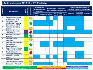
Download
1 / 1
10 likes | 139 Views
Comparative Diffusion Tensor Imaging (DTI) Study of Tool Use Pathways in Humans, Apes and Monkeys. Ashwin G. Ramayya 1,2 , Matthew F. Glasser 1 , David A. Gutman 5 , James K. Rilling 1,3,4,5

E N D
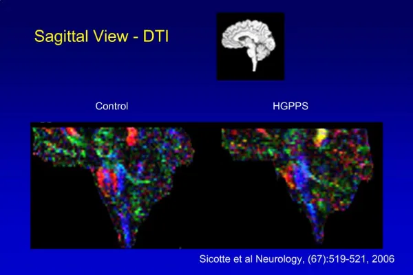
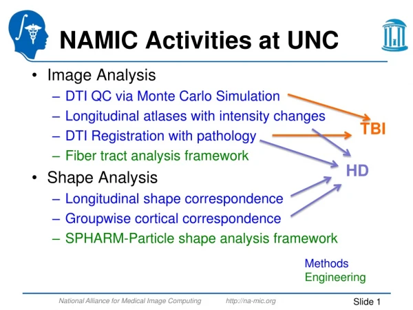
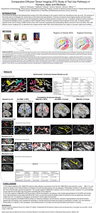
Comparative Diffusion Tensor Imaging (DTI) Study of Tool Use Pathways in Humans, Apes and Monkeys Ashwin G. Ramayya1,2, Matthew F. Glasser1, David A. Gutman5, James K. Rilling1,3,4,5 1Department of Anthropology, 2Neuroscience and Behavioral Biology Program, 3Center for Behavioral Neuroscience, 4Yerkes National Primate Research Center, 5Department of Psychiatry and Behavioral Sciences, Emory University INTRODUCTION One behavioral attribute that distinguishes humans from other primates is the extent to which we manufacture and use tools. The purpose of this study was to investigate the neural basis of this behavioral specialization. Previous functional neuroimaging studies and brain lesion cases have implicated a network of four left hemisphere regions as being central to human tool use: 1) posterior temporal cortex involved in conceptual knowledge of tools, 2) posterior inferior parietal cortex involved in acquired motor programs for tool use, 3) anterior inferior parietal cortex and 4) inferior middle frontal cortex, both involved in grasping and manipulating objects (Johnson-Frey et al 2005). We used diffusion tensor imaging (DTI) to describe and compare white matter fiber tracts linking these four regions in humans, apes and monkeys. METHODS Regions of Interest (ROI) Segment Summary Rhesus macaque n=2 Bonobo n=2 Chimpanzee n=4 Human n=32 ~2-3 mya ~8-10 mya ~30 mya IMAGE ACQUISITION: Post-mortem DTI scans were acquired from three common chimpanzee, two pygmy chimpanzee (bonobo) and two rhesus macaque specimens using a spin echo sequence. In vivo DTI scans were acquired from thirty-two humans (age 18-50) and one adult male chimpanzee using single shot and segmented EPI protocols, respectively. Spatial resolution of human, ape and monkey scans were 1.7 x 1.7 x 2.0 mm, 1.5 mm isotropic and 0.55 mm isotropic, respectively. All non-human primate scans and thirteen human scans were acquired with 60 diffusion directions. The remaining nineteen human scans were acquired with 12 directions. IMAGE ANALYSIS: For each scan, cortical surface landmarks were used to define three anatomical regions of interest (ROI) (see figure above): 1) middle temporal gyrus (MTG in red), 2) anterior supramarginal gyrus (Ant SMG in blue) and 3) posterior supramarginal/angular gyrus (SMG/AG in pink). Frontal lobe projections from AntSMG and SMG/AG to the frontal lobe were used to define a fourth ROI in the frontal lobe (Frontal in yellow). In the macaques, the two parietal ROI were combined due to the lack of an upper projection of the sylvian fissure. Four pathways were tracked and compared across hemispheres and species: 1) MTG AntSMG (blue), 2) MTG SMG/AG (pink), and 3) AntSMG Frontal (yellow) 4) SMG/AG Frontal (orange). The 12 direction images were analyzed with the Siemens DTI Task Card software (MGH), which uses a deterministic tractography algorithm. The remaining data was analyzed using FSL using a two-fiber direction probabilistic algorithm. The 60-direction human data were spatially normalized to standard space, to illustrate group average tractography results. RESULTS Deterministic 12-direction Human Results (n=19) Representative Subject (RX): *AI= (# samples in the left hemisphere) – (# samples the right hemisphere) (total # in both hemispheres) Probabilistic 60-direction Results Humans (n=13) Ant.SMG MTG SMG/AG MTG Ant.SMG Frontal SMG/AG Frontal Representative Subject (OCMR_04): Chimpanzees (n=4) Representative Subject (Artifee): Bonobos (n=2) Representative Subject (Suke): Macaques (n=2) Representative Subject (Macaque_2): Absent Summary: CONCLUSIONS 1) The yellow segment (Ant. SMGFrontal) provides afferent connections from the Ant. SMG ROI to the premotor cortex (BA 4, 6), and allows for spatio-motor transformations necessary for goal-directed actions such as grasping. Predictably, it present in all species, and is the most evolutionarily conserved segment. The orange segment (SMG/AG) is likely to have a similar function, however it is unremarkable across species as the ROI may have been excluding part of the segment. 2) The pink segment (SMG/AG MTG) provides a direct link between conceptual, multi-modal representations of the MTG and stored motor representations associated with BA 40/39. Such connectivity would be ideally suited for behaviors such as gesturing, and some simple tool-use. It is absent in macaques, but present in all the apes and humans 3) The blue segment (Ant.SMG MTG) allows for interaction between ventral-stream, semantic representations and dorsal stream, spatiomotor processing. Such anatomical synthesis of ventral and dorsal stream processes is essential to complex -tool use behavior. Reasonably, it is absent in the macaques, only weakly present in the apes, but strong and highly asymmetric in humans REFERENCES Johnson-Frey SH, Newman-Norlund R, Grafton ST (2005): A distributed left hemisphere network active during planning of everyday tool use skills. Cereb Cortex 15:681-695. Grant suport was provided by the Emory University Research Committee and the Center for Behavioral Neuroscience, Atlanta, GA.