Diffusion Weighted Imaging Tensor Analysis
Diffusion Weighted Imaging Tensor Analysis. Vincent A. Magnotta Associate Professor March 21, 2011. Diffusion Tensor Analysis Flow Chart. DTIPrep 1. Verify Acquisition 2. Artifact Detection 3. Motion Correction 4. Update Gradient Directions 5. Remove Bad Data. Images Format


Diffusion Weighted Imaging Tensor Analysis
E N D
Presentation Transcript
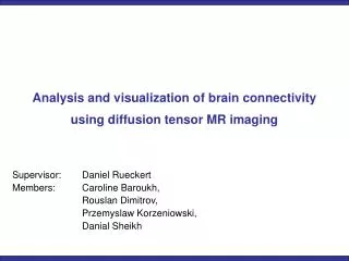
Diffusion Weighted ImagingTensor Analysis Vincent A. Magnotta Associate Professor March 21, 2011
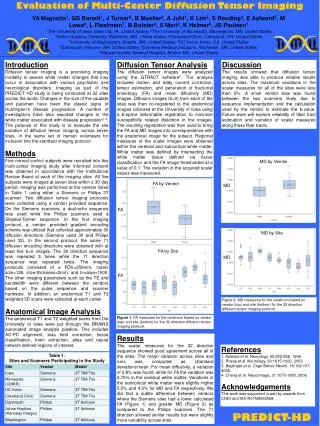
Diffusion Tensor Analysis Flow Chart DTIPrep 1. Verify Acquisition 2. Artifact Detection 3. Motion Correction 4. Update Gradient Directions 5. Remove Bad Data Images Format Conversion DTI Data Collection (DICOM) Concatenate Data Extract B0 Image Resample Images Into ACPC Space Non-Rigid Co-Register With AC-PC Aligned T1 Rigid Co-Register With AC-PC Aligned T1 Create Diffusion Scalar Images Generation Of Diffusion Tensor
Diffusion Tensor Image Analysis • Image format conversion • Change from DICOM to Nifti or NRRD image formats • Rotate applied diffusion gradients • Motion Correction • Account for patient motion and eddy current artifacts • Generation of Diffusion Tensor • Includes possible edge preserving low pass spatial filtering • Use rotated diffusion directions • Create Diffusion Tensor scalar maps • Mean diffusivity • Fractional Anisotropy • Relative anisotropy • Radial Diffusivity • Axial Diffusivity • Co-register with anatomical image • Rigid • Non-Rigid (B-Spline)
Image Format Conversion • Convert from DICOM to NRRD format • Nearly Raw Raster Data • Defines origin, spacing, orientation, Diffusion Gradients, and Measurement Frame • Coordinate frame for the applied diffusion gradients • All information obtained from DICOM header • Siemens, Philips, and GE scanners DicomToNrrdConverter \ --inputDicomDirectory /home/vince/images/dti_images \ --outputDirectory /home/vince/images \ --outputVolume /home/vince/images/SUBJECT_DWI.nhdr
DTI Concatenation • Concatenate multiple DTI runs together • Improve SNR of tensor estimation • Runs can contain any number of gradient directions and orientations gtractConcatDwi --outputVolumedti.nhdr\ --inputVolumedti_parta.nhdr,dti_partb.nhdr
Improving DTI measures: DTIPrep • From UNC, Zhexing Liu • Purpose of DTIPrep: provide individual and group quality control of DWI/DTI data sets in GUI and command line mode • Detect and remove artifacts that often appear in DWI data • Prevent artifacts from creating DTI estimation errors in tensor principle orientation (premature fiber tracking termination) and scalars • Prevent low consistency in quality control associated with current visual checking of DWI data sets
DTIPrep: Quality Control Pipeline • Image information checking • Diffusion information checking • Slice-wise intensity artifact checking • Interlace-wise venetian blind artifact checking • Baseline averaging • Eddy-current and head motion artifact correction • Gradient-wise checking (motion artifact checking)
DTIPrep: Quality Control Pipeline • Image information checking • Image space • Image directions • Image size • Image spacing • Image origin • Cropping • Diffusion information checking • b value • Diffusion gradient vectors • Tolerance tests • Replacement of diffusion gradient vectors with those in acquisition protocol
DTIPrep: Quality Control Pipeline • Venetian blind artifact detection • Baseline averaging • Motion between baseline scans is removed by rigidly registering all baseline scans and averaging them together • The averaged baseline image is used as a reference for subsequent eddy-current and head motion artifact correction for all gradients • Eddy-current and head motion artifacts correction • Resulting image is SUBJECT_DWI_Qced.nhdr DTIPrep –DWINrrdFile /home/vince/images/SUBJECT_DWI.nhdr \ --xmlProtocol /home/vince/images/default.xml \ --default --check --outputFolder/home/vince/images
DTIPrep Outputs • NRRD file containing • Single baseline average image (motion corrected) • Corrected Diffusion gradients • Passed quality control (slice-wise & interlace) • Head motion corrected (Rigid register to baseline with gradient direction adjustments relative to anatomical frame of reference) • Eddy current corrected (Affine register to baseline) • SUBJECT_DWI_Qced.nhdr • Report on excluded diffusion gradients • SUBJECT_DWI_QcReport.txt • Optional outputs: NRRD files of excluded diffusion gradients from each quality control step • DTIPrep outputs GTRACT
DTIPrep: Quality Control Pipeline • 2.4 Slice-wise intensity related artifacts checking Slice-to-slice correlation value Analysis region Slice number We propose to use Normalized Correlation (NC) between successive slices across all the diffusion gradients for screening the intensity related artifacts.
DTIPrep: Quality Control Pipeline • 2.5 Interlace-wise Venetian blind artifact checking Translation (mm), Angle of rotation (degrees) Gradient number Venetian blind like artifacts can be detected via correlations and motion parameters between the interleaved parts for each gradient volume.
DTIPrep: Quality Control Pipeline • 2.8 Gradient-wise checking Motion artifact residuals after eddy-current and head motion corrections can be detected via motion parameters between baseline and each of the gradients.
DTIPrep Impact on FA values • Exclusion of optimal number of gradients minimized the standard deviation in FA values • Standard deviation was lowered in a single scan processed by DTIPrep Standard deviation in FA Standard deviation in FA 26% 27% 9% 21% Without DTIPrep With DTIPrep
Create Diffusion Tensor • Create Tensor representation of diffusion process • Defined by 6 unique parameters • Allows for edge preserving low pass filtering (median) whose radius is defined in voxels • Removal of background signal gtractTensor \ --inputVolumeSubject_DTIPREP.nhdr \ --outputVolumeSUBJECT_Tensor.nhdr \ --medianFilterSize1,1,1 --backgroundSuppressingThreshold50 --b0Index 0
Rotationally Invariant Scalar Generation • Eigen analysis of tensor data • Creates a variety of scalars: • FA – Fractional Anisotropy • MD – Mean Diffusivity • RA – Relative Anisotropy • LI – Lattice Index • AD – Axial Diffusivity • RD – Radial Diffusivity gtractAnisotropyMap \ --inputTensorVolumeSubject_Tensor.nhdr \ --outputVolumeSUBJECT_FA.nii.gz \ --anisotropyType FA
Image Extraction and Clipping • Extract B0 image • Clip B0 image to remove skull using AFNI extractNrrdVectorIndex --index 0\ --inputVolumeSubject_DTIPREP.nhdr \ --outputVolume Subject_B0.nii.gz 3dAutomask -prefix Subject_DWI_B0_mask.nii.gz \ Subject_B0.nii.gz 3dcalc -a Subject_DWI_B0_mask.nii.gz \ -datum short -expr "a*1" \ -prefix Subject_B0_maskShort.nii.gz 3dcalc -a Subject_B0_maskShort.nii.gz \ -b Subject_DWI_B0.nii.gz -expr "a*b" \ -prefix Subject_DWI_B0_Brain.nii.gz
DWI to Anatomical Registration • Utilize BRAINSFit image registration • Supports Mutual Information registration metric • Non-linear image registration • B-splines can be used to correct for susceptibility artifacts • Eliminates the need for field maps BRAINSFit –movingVolume Subject_DWI_B0_Brain.nii.gz \ --fixedVolume Subject_clippedT1.nii.gz\ --transformTypeRigid,BSpline \ --numberOfSamples 500000 \ --splineGridSize 12,12,12 \ --outputTransformSUBJECT_ACPC.mat \ --initializeTransformModeuseMomentsAlign
Invert Transform • Provide a mapping from AC-PC apace back to the DTI space • Approximate inverse is computed using Thin Plate Spline (TPS) transforms • Used to map ROIs into DTI space for fiber tracking gtractInvertBSplineTransform \ --inputTransformSUBJECT_ACPC.mat \ --outputTransformSUBJECT_ACPC_Inverse.mat \ --inputReferenceVolume Subject_clippedT1.nii.gz
Resample DTI Scalars • Place rotationally invariant scalars into the space of anatomical images • Resample B0 image to check quality of registration BRAINSResample \ --referenceVolumeSUBJECT_T1.nii.gz \ --inputVolumeSUBJECT_FA.nii.gz \ --warpTransformSUBJECT_ACPC.mat\ --outputVolumeSUBJECT_FA_ACPC.nii.gz --interpolationMode Linear
Diffusion Tensor Scalar Measurements • Lobar Talairach Analysis • Frontal, Temporal, Parietal, Occipital, and Cerebellar white matter measurements • White matter region defined using both FA and tissue classified images • BRAINS measurement script exists
Diffusion Tensor Scalar Measurements A-P • Analysis of Anisotropy from Anterior-Posterior based on Talairach Atlas • Divide regions from A-D and F-I in half • Retain sizes of E1, E2 and E3 • BRAINS script exists
SPM Analysis • Co-register to Atlas image • Apply transform to DTI scalar image • Smooth Scalar images • Threshold to White matter regions • Possible issues with anatomic variability
Base on the directional information provided by DTI, fiber tracking can be used to explore the underlying white matter fiber structure non-invasively Fiber Tracking - Introduction