Flat panels detectors (FPDs)
350 likes | 566 Views
Flat panels detectors (FPDs). I.I. vs. FPD. Low weight and dimension ). Pros of FPDs. Stability through device life. Better image quality. Non ideal trajectory. Irregular sampling step. Distorsion in C-Arm and correction. Off-line calibration required.

Flat panels detectors (FPDs)
E N D
Presentation Transcript
Flat panels detectors (FPDs) I.I. vs. FPD Low weight and dimension) ProsofFPDs Stability through device life Better image quality
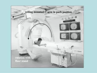
Non ideal trajectory Irregular sampling step Distorsion in C-Arm and correction Off-line calibrationrequired A calibration ring phantom with metal beads is used for calibration. Calibration should be performed for each used scan trajectory, since mechanical distortion is specific Calibration sets the correct correspondence between 2D projection and 3D position. It is repeated weekly.
3D Angiography Workstation Processing SSD (Surface Shaded Display) MIP (Maximum Intensity Projection)
3D Angiography Workstation Processing Surface vs. Volume Rendering Virtual Navigator
3D Angiography Workstation Processing Processing della workstation Coils and Clips Opposite to DSA (Digital Subtraction Angiography), which deletes still objects, this algorithm does reconstruct high contrast objects not moving and varying during the scan and enhances them over the vascular anatomy Calibrated anatomycal measures
3D Angiography Risultati Immagine DSA standard Aneurismi multipli dell’arteria carotide interna Viste ottenute con Volume Rendering (differenti proiezioni) Aneurismi multipli dell’arteria carotide interna
Multislice helical CT Giuseppe Gentil, Francesco Maffesanti, Massimo Molinari, 2005/06
direction ofcontinuousbed shift multi-row detector array trajectory of the rotatingx-ray tube focal spot scan start
z axis multi-row detector array
CRANIOPLASTICS • Application in the field of maxillo-facial surgery • Precise cranial CT scan • Import into specific SW environment; e.g., Mimics • (Materialize’s InteractiveMedicalImage • Control System) • Segmentation of skull • Vectorization into a CAD environment • Design of prosthesis • Construction by 3D rapid prototiping GANTRY TILT = 0°
Segmentation by Region Growing and regular contouring by NURBS Polinomial contours by NonUniform Rational B-Splines (NURBS) 3D
Application: from 3D virtual model to real model by rapid prototyping
The polymer prototype is not directly used. Conversely, it is used as 3D model for fabrication or to create a mold (it stampo) were the final material is cast . Final material can be autologous or donor bone, hydroxyapatite, PMMA, Titanium silicon-rubber mold
I Materiali I materiali usati finora per la cranioplastica sono stati molteplici, ma molti di loro sono stati abbandonati per diversi motivi: scarsa biocompatibilità, ridotta maneggevolezza nel modellamento, alto costo… I materiali maggiormente utilizzati oggi sono: Osso autologo Titanio Idrossiapatite PMMA La Cranioplastica
Tecnica Chirurgica • Posizionamento paziente • Rimozione adesione pelle–dura madre La Cranioplastica
3. Applicazione acqua ossigenata (disinfettare e coagulare) La Cranioplastica