Download
1 / 60
600 likes | 721 Views
Learn about sex determination, gonad development, gametogenesis, and differences between male and female reproductive systems. Explore the stages of cell division in sperm and egg development.

E N D
Sex Determination • Dimorphism- physical differences in females and males other and reproductive organs. • Physically distinct-breast, hips, muscle mass, areas of body fat storage, etc. • Gonads produce gametes (eggs and sperm) • Male gonads testes sperm • Female gonads ovaries eggs • Internal and external genitalia – glands and ducts that connect the gonads to the external genitalia
Sex Determination • Chromosomes – a DNA segment that holds genes. Chromosomes come in pairs called homologous pairs • Autosomes – code for body characteristics but do not determine sex • 22 pairs • Sex Chromosomes -X & Y • men are XY women are XX Figure 26-1
Sex Determination Inheritance of X and Y chromosomes Each parent contributes ½ the genome to an offspring. During sex determineation; mothers always contribute the X chromosome. Father’s have a 50:50 chance of giving an X or Y Figure 26-2
Sexual Differentiation • Bipotential tissues – undifferentiated tissues the can develop into male or female reproductive structures. • SRY gene – sex-determining region of the Y chromosome that will guides the development of male genitals. • Gonad - the reproductive organ that produces the gamete • Testis or ovary – primary reproductive organ, all other reproductive structures are accessory structures that make fertilization and pregnancy possible. • Wolffian duct- derived from the embryonic kidney, it develops into the vas deference once the testes release Anti-Mullerian hormone. • Mullerian duct - derived from the embryonic uterus, it develops into the fallopian tube while Wolfian duct disappear in the absence of testorone.
Development of Internal Organs Figure 23-3a
Development of External Genitalia Figure 26-3b
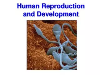
Gametogenesis • Gamete production occurs in primary sex organs. Begins in utero, stops during childhood, resumes in puberty and continues through adulthood. The process is timed differently in males & females • Meiosis- cell division occurring only in gametes that yields four haploid cells (different from mitosis) • Primary gamete-produced at completion of Meiosis I. The cell is genetically diverse, haploid (no chromosome pairs) and with sister chromatids (copies= 46 total) • Secondary gamete-produced at completion of Meiosis II. The cell is genetically diverse, haploid, and with no sister chromatids (23 total) • Haploid gamete- carries half the genome, matures into a viable cells that participates in fertilization
Comparing Mitosis and Meiosis Mitosis Meiosis • Mitosis yields two identical diploid cells. It’s happens in all body cells (somatic) • Meiosis yields four genetically diverse haploid cells. It only happens in sex cells (gametes) Meiosis I Parent cell (2n) Parent cell (2n) Chromosome duplication Chromosome duplication Tetrad Crossing over Daughter cells 2n 2n Meiosis II Visual Summary 8.3 n n n n Daughter cells
Gametogenisis in the semineferous tubules of the testes. Stem cells first go through mitosis & then meisosis
Spermatogenesis STAGE OF CELL DIVISION MALE FEMALE Spermatogonium MITOSIS Germ cell proliferation 46 chromosomes per cell (only two shown here) Embryo Embryo 46 (diploid) Spermatogonia MEIOSIS DNA replicates but no cell division occurs. Primary spermatocyte Sister chromatids 2 sets of 46 chromosomes Reproductive adult First meiotic division Secondary spermatocyte Primary gamete divides into two secondary gametes. 23 chromosomes duplicated Reproductive adult Second meiotic division Spermatids develop into Secondary gamete divides. 23 chromosomes (haploid) Sperm One primary spermatocyte yields 4 sperm. In males meiosis begins in adulthood after puberty, all cells generated by meiosis can develop into sperm. Figure 26-5 (4 of 9)
Spermatozoa Structure • Head • Acrosome and nucleus • Midpiece • Centrioles and mitochondria • Tail (flagellum) • Microtubules
Oogenesis In females stem cells go through mitosis and creates all the oocytes that could mature into an ovum. Meiosis begins before birth, is arrested,and then resumes after puberty but is not completed until fertilization occurs.
Gametogenesis & Fertilization STAGE OF CELL DIVISION MALE FEMALE Spermatogonium Oögonium MITOSIS Germ cell proliferation 46 chromosomes per cell (only two shown here) Embryo Embryo 46 (diploid) Oögonia Spermatogonia MEIOSIS DNA replicates but no cell division occurs. Primary spermatocyte Primary oocyte Sister chromatids Sister chromatids 2 sets of 46 chromosomes Reproductive adult First meiotic division First polar body Secondary oocyte (egg) Secondary spermatocyte Primary gamete divides into two secondary gametes. 23 chromosomes duplicated (may not occur) Reproductive adult Second meiotic Disintegrates division Spermatids develop into Secondary gamete divides. Egg released from ovary at ovulation 23 chromosomes (haploid) Sperm One primary spermatocyte yields 4 sperm. One primary oocyte yields 1 egg. FERTILIZATION Second polar body disintegrates. Unfertilized egg passes out of body. Zygote Figure 26-5 (9 of 9)
Synthesis Pathways of Steroid Hormones • Steroid hormones contain cholesterol, are structurally similar and share production pathways with other steroid hormones • Ovary • Progesterone • Estrogen • Testis • Testosterone
Regulation of Reproduction by Endocrine System • Both males and females produce the following, the primary target tissues are the gonads. The gonads produce androgens (predominate in males) and estrogens (predominate in females). • Hypothalamus: pulse generator of • Gonadotropin releasing hormone (GnRH) • Anterior Pituitary • Lutenizing hormone (LH) • Follicle stimulating hormone (FSH) • Inhibins and activins- influence the secretion of FSH
General Pathways Internal and environmental stimuli CNS GnRH Hypothalamus Short-loop negative feedback KEY Stimulus Anterior pituitary Integrating center Efferent pathway Effector Long-loop feedback may be negative or positive Tissue response LH FSH Gonads (ovaries or testes) Females only Endocrine cells Gamete production Steroid and peptide hormones General pattern of hormonal control of reproduction • Long-loop: hormone production by gonads alters • GnRH • FSH • LH • Short-loop: feedback from pituitary alters • GnRH Figure 26-7
Histology of the Testes • Seminiferous tubules contain • Spermatogonia- goes through various stages to become sperm (spermatogonia, spermatocyte, spermatids, & sperm) • Sertoli cells (sustentacular cells)- found between spermatogonia, form part of the blood-testes barrier produce inhibin & activin • Interstitial tissue (found outside of seminiferous tubules but within testicles) • Leydig cells- produce testosterone, active in fetus and after puberty
Regulation of Spermatogenesis KEY GnRH Hypothalamus Integrating center Efferent pathway Effector Tissue response Anterior pituitary FSH LH Spermatogonium Inhibin Spermatocyte Testes Sertoli cell Second messenger Cell products Sertoli cell Androgen-binding protein (ABP) ABP T • GnRH FSH Sertoli cells spermatocyte maturation • Inhibin feedback • FSH, testosterone • Short and long loops
Regulation of Spermatogenesis • GnRH LH Leydig cells testosterone sex characteristics • Inhibin feedback • FSH, testosterone • Short and long loops
Male Reproductive Structures • Male reproductive structures are designed to create sperm, deliver it internally, and provide it with chemicals to allow it to survive outside the body
Male Reproductive Structures Figure 26-9b
Male Reproductive Structures Accessory glands: produce secretions that in addition to sperm, form semen. 1. Seminal vesicles: secrete a fluid that contains fructose (sugar), nutrients, prostagladins to stimulate the urethra to contract, substances that suppress the immune system against sperm in females, enzymes the enhance sperm mobility, and enzymes that thicken the ejaculate. 2. Prostate gland: release a milky white fluid that enhances sperm mobility & thickens ejaculate. Susceptible to tumors & STDs. 3. Bulbourethral glands: secrete a mucus for lubrication and neutralizes acid in urethra. Figure 26-8 (1 of 2)
Female Reproductive Structures • External genitalia • Vulva • Labia Majora & Minora • Clitoris • Urethral & Vaginal opennings • Passageway • Urethra • Vagina • Hymen • Birth canal
Female Reproductive Structures The female reproductive system is designed for ova(egg) production, fertilization, fetal development, and fetal delivery. It involves a uterine and ovarian cycle
Female Reproductive Structures • These structures are supported by the broad ligament. The uterus is not the place of fertilization, thus changes to the lining of the uterus occur in preparation of implantation and then development of the fetus
Ovaries: Structures • Follicle: primordial, primary, secondary, matured (antrum), ruptured • Oocyte • Follicular cells • Thecal cells • Granulosa cells • Corpus luteum • Corups albicans
Phases of the Ovarian & Uterine Cycle • Primary hormones • GnRH from the hypothalamus • FSH and LH from the anterior pituitary • Estrogen, progesterone, and inhibin from the ovary • Ovarian Cycle: • Follicular phase: Follicle growth in ovary (egg matures) • Ovulation: Ripened follicles and release of oocyte(s) • Luteal phase: Ruptured follicle becomes corpus luteum in preparation for pregnancy • Uterine Cycle: • Menses: No pregnancy, Bleeding from uterus • Proliferative phase: New layer of endometrium in preparation of pregnancy • Secretory phase: Conversion of endometrium to secretory structure
Hormonal Control of the Menstrual Cycle:Follicular Phase and Ovulation (a) Early to mid-follicular phase (b) Late follicular phase and ovulation GnRH GnRH Hypothalamus Pituitary FSH LH LH FSH Follicle Follicle Thecal cells Granulosa cells Granulosa cells Thecal cells Androgens Inhibin Androgens High estrogen output Small amount of progesterone Estrogens LH FSH KEY Ovum Stimulus Integrating center Follicle Corpus luteum Efferent pathway Inhibin Progesterone Estrogen Tissue response Figure 26-14 (2 of 4)
The Menstrual Cycle: Ovarian & Uterine Cycle Phases of the Ovarian Cycle Follicular Phase Gonadotrophic hormone levels FSH LH Ovarian cycle Primary follicle Theca Ovarian hormone levels Estrogen Inhibin Progesterone Uterine cycle Phases of the Uterine Cycle MENSES 36.7 Basal body temperature (˚C) 36.4 DAYS 28/0 7 14 21 28/0 • FSH stimulates follicular development • Maturation to secondary and tertiary follicle • Granulosa cells produce estrogen • Positive feedback limits more follicles • Negative feedback decreases FSH and LH secretion • LH stimulates thecal cells to produce androgens
The Menstrual Cycle: Ovarian & Uterine Cycle Phases of the Ovarian Cycle Follicular Phase Luteal Phase Gonadotrophic hormone levels FSH LH Ovarian cycle Primary follicle Theca Antrum Ovarian hormone levels Estrogen Inhibin Progesterone Uterine cycle Phases of the Uterine Cycle PROLIFERATIVE PHASE MENSES 36.7 Basal body temperature (˚C) 36.4 DAYS 28/0 7 14 21 28/0 • High levels of estrogen • LH surge and FSH spike • Egg release • High levels of inhibin • Inhibits production of FSH • Decrease new follicle development • Low levels of progesterone • Positive feedback • GnRH and LH
Hormonal Control of the Menstrual Cycle: Early to Mid-Luteal Phase (a) Early to mid-follicular phase (b) Late follicular phase and ovulation (c) Early to mid-luteal phase GnRH GnRH GnRH Hypothalamus Pituitary FSH LH LH FSH FSH LH Follicle Follicle Corpus luteum (from ovulated follicle) Thecal cells Granulosa cells Granulosa cells Thecal cells secretes Androgens Inhibin Estrogen Androgens Progesterone High estrogen output Small amount of progesterone Inhibin Estrogens LH FSH KEY Ovum Stimulus Integrating center Follicle Corpus luteum Efferent pathway Inhibin Progesterone Estrogen Tissue response Figure 26-14 (3 of 4)
The Menstrual Cycle: Ovarian & Uterine Cycle Phases of the Ovarian Cycle Follicular Phase Luteal Phase Gonadotrophic hormone levels FSH LH Ovarian cycle Primary follicle Corpus luteum formation Theca Ovulation Antrum Ovarian hormone levels Estrogen Inhibin Progesterone Uterine cycle Phases of the Uterine Cycle PROLIFERATIVE PHASE MENSES SECRETORY PHASE 36.7 Basal body temperature (˚C) 36.4 DAYS 28/0 7 14 21 28/0 • Granulosa cells • Form corpus luteum • Secretesprogesterone • High levels of progesterone and estrogen maintain endometrium • Inhibin continues to limit new follicular development
Hormonal Control of the Menstrual Cycle: Late Luteal Phase (a) Early to mid-follicular phase (b) Late follicular phase and ovulation (c) Early to mid-luteal phase (d) Late luteal phase GnRH GnRH GnRH GnRH Hypothalamus Tonic secretion resumes Pituitary FSH LH FSH LH LH FSH FSH LH Corpus luteum dies Follicle New follicles begin to develop Follicle Corpus luteum (from ovulated follicle) Thecal cells Granulosa cells Granulosa cells Thecal cells secretes Androgens Inhibin Estrogen and progesterone Estrogen Androgens Progesterone High estrogen output Small amount of progesterone Inhibin Estrogens LH FSH KEY Ovum Stimulus Integrating center Follicle Corpus luteum Efferent pathway Inhibin Progesterone Estrogen Tissue response Figure 26-14 (4 of 4)
The Menstrual Cycle: Ovarian & Uterine Cycle Phases of the Ovarian Cycle Follicular Phase Luteal Phase Gonadotrophic hormone levels FSH LH Ovarian cycle Primary follicle Corpus luteum formation Mature corpus luteum Corpus albicans Theca Ovulation Antrum Ovarian hormone levels Estrogen Inhibin Progesterone Uterine cycle Phases of the Uterine Cycle PROLIFERATIVE PHASE MENSES SECRETORY PHASE 36.7 Basal body temperature (˚C) 36.4 DAYS 28/0 7 14 21 28/0 • Pregnancy • Maintains high levels ofprogesterone, estrogen, and inhibin • No pregnancy • Decreased levels of progesterone, estrogen, and inhibin • Menses • High levels of FSH and LH • New follicle development
Procreation: Sexual Response • Phases of coitus (sexual intercourse or copulation) • Excitement • Plateau • Orgasm • Resolution • Erection reflex • CNS and spinal integration • Emission • Ejaculation • Erectile dysfunction (ED)
Erection and Ejaculation in Males KEY Stimulus Receptor Afferent pathway Integrating center Efferent pathway Effector Tissue response Parasympathetic stimulated Sympathetic inhibited Penile arterioles vasodilate. Spinal cord Penis Erection Tactile stimuli Sensory neuron Mechanoreceptor Figure 26-15 (5 of 7)
Erection and Ejaculation in Males Thoughts of Sex!! KEY Stimulus Receptor Afferent pathway Higher brain centers Erotic stimuli Integrating center Efferent pathway Effector Tissue response Ascending sensory pathway Parasympathetic stimulated Sympathetic inhibited Penile arterioles vasodilate. Spinal cord Penis Erection Tactile stimuli Sensory neuron Mechanoreceptor Figure 26-15 (6 of 7)
Erection and Ejaculation in Males Thoughts of Sex!! KEY Stimulus Receptor Afferent pathway Higher brain centers Erotic stimuli Integrating center Efferent pathway Effector Tissue response Descending autonomic pathways Ascending sensory pathway Parasympathetic stimulated Sympathetic inhibited Penile arterioles vasodilate. Spinal cord Penis Erection Tactile stimuli Sensory neuron Mechanoreceptor Figure 26-15 (7 of 7)
Pregnancy Prevention • Contraceptives • Abstinence-also prevents STDs • Barriers-prevent sperm from entering cervix • Surgical- prevent the entry of gametes • Pills- affect uterine lining and ovulation http://portal.stii.dost.gov.ph/sntpost/frames/OcttoDec03/grapix/contraceptives.jpg
Fertilization • Sperm capacitation • Swimming and attractants • Egg contact • Penetration • Acrosomal reaction • Nuclear fusion • Cortical reaction • Zygote
Fertilization Figure 26-16a
Fertilization • When fertilization occurs the sperm does not enter the egg but the fusion of the membranes causes the removal of receptors on the egg so no other sperm can fuse. Notice that meiosis II is only completed if the egg is fertilized
Zygote Development: Ovulation, Fertilization, and Implantation Days 2-4: Cell division takes place. Day 1: Fertilization Days 4-5: Blastocyst reaches uterus. 3 2 4 Inner cell Zygote Fallopian tube Egg Blastocyst Ovary 1 Ovulation 5 Days 5-9: Blastocyst implants. Uterus Figure 26-18, steps 1–5
Further Embryonic Development • Chorion and amnion • Placenta • Exchange site • Hormones • Human chorionic gonadotropin (hCG) • Human placental lactogen (hPL) • Estrogen and progesterone
The Placenta Figure 26-19a