Download
1 / 15
150 likes | 348 Views
Clinical features: pulmonary disease. Pulmonary Disease symptoms Classically include cough, fever, and sweating.

E N D
Pulmonary Disease symptoms Classically include cough, fever, and sweating. • Coughis nearly universal; typically, it is initially dry but then progresses with increasing volumes of purulent secretions and the variable appearance of blood streaking or gross hemoptysis. • Feverishnessis common as the disease advances. • Sweating, including drenching night sweats, is typical. • Other common complaints include: fatigue, weight loss, nonpleuritic chest pain, and dyspnea. signs • Feverwith peaks as high as 40 to 41° C, typically occurring in the evening. • Localized ralesare early findings, • Wheezing or regionally diminished breath sounds, or both, may be heard in patients with peribronchial or endobronchial airway narrowing..
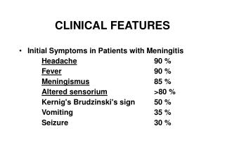
Clinical setting Primary pulmonary TB (=infection of a previously uninfected (tuberculin-negative individual) • Most patients develop a self-limiting febrile illness, usually is manifested only by development of a positive tuberculin skin test. • Occasionally, the patient develops sufficient symptoms of fever and nonproductive cough with chest radiographic finding of patchy or lobular infiltrates, often with associated hilaradenopathy. Progressive primary disease (=Early progression of infection to disease) • May appear during the course of the initial illness or after a latent period of weeks or months. • May manifest as miliary tuberculosis, sometimes with meningitis, or as pulmonary disease of the apical and posterior segments of the upper lobes or lower lobe disease.
Miliary TB = Blood-borne dissemination • May present acutely, but more frequently is characterised by 2-3 weeks of fever, night sweats, anorexia, weight loss and a dry cough. • Hepatosplenomegaly may be present and the presence of a headache may indicate co-existent tuberculous meningitis. • Auscultation of the chest is frequently normal, although with more advanced disease widespread crackles are evident. • Fundoscopy may show choroidal tubercles. • The classical appearances on chest X-ray are those of fine 1-2 mm lesions ('millet seed') distributed throughout the lung fields, although occasionally the appearances are coarser. • Anaemia and leucopenia may be present • Negative tuberculin skin test in 50% of pateints. • Confirmation ; by biopsy (granulomas and/or acid-fast bacilli demonstrated) of liver or bone marrow
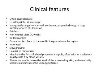
Post-primary pulmonary TB = exogenous ('new' infection) or endogenous (reactivation of a dormant primary lesion) infection in a person who has been sensitized by earlier exposure. • It is the most frequent presentation and characteristically occurs in the apex of an upper lobe where the oxygen tension favours survival of the strictly aerobic organism. • The onset is usually insidious, developing slowly over several weeks. Systemic symptoms accompanied by progressive pulmonary symptoms . • Radiological changes include ill-defined opacification in one or both of the upper lobes, and as progression occurs, consolidation, collapse and cavitation develop to varying degrees . • In extensive disease, collapse may be marked and result in significant displacement of the trachea and mediastinum. Occasionally, a caseous lymph node may drain into an adjoining bronchus resulting in tuberculous pneumonia.
Diagnosis • The presence of an otherwise unexplained cough for more than 2-3 weeks, particularly in an area where TB is highly prevalent, or typical chest X-ray changes should prompt further investigation. • Direct microscopy of sputum is the most important first step. The probability of detecting acid-fast bacilli is proportional to the bacillary burden in the sputum • The most effective techniques are the Ziehl-Neelsen and rhodamine-auramine stains. The latter causes the tuberculous bacilli to fluoresce against a dark background and is easier to use when numerous specimens need to be examined; • A positive smear is sufficient for the presumptive diagnosis of TB but definitive diagnosis requires culture. • Smear-negative sputum should also be cultured, as only 10-100 viable organisms are required for sputum to be culture-positive.
Mycobacteria TB grow slowly and may take between 4 and 6 weeks to appear on solid medium such as Löwenstein-Jensen or Middlebrook. • Faster growth (1-3 weeks) occurs in liquid media such as the radioactive BACTEC system • The BACTEC method is commonly used in developed nations and detects mycobacterial growth by measuring the liberation of 14CO2, following metabolism of 14C-labelled substrate present in the medium. !!!!!!!!!!!!!!!!!!!!!!!!!!!!!!!!!!!!!!!!!!!!!!!!!!!!!!!!!!!!!!!!!!!!!!!!!!!!!! • New strategies for the rapid confirmation of TB at low cost are being developed; these include the nucleic acid amplification test (NAT), designed to amplify nucleic acid regions specific to Mycobacteria TB • Drug sensitivity testing is particularly important in those with a previous history of TB, treatment failure or chronic disease, those who are resident in or have visited an area of high prevalence of resistance, or those who are HIV-positive. • The detection of rifampicin resistance, using molecular tools to test for the presence of the rpo gene currently associated with around 95% of rifampicin-resistant cases, is important as the drug forms the cornerstone of 6-month chemotherapy.
Chronic complications of pulmonary TB Pulmonary • Massive haemoptysis • Corpulmonale • Fibrosis/emphysema • Atypical mycobacterial infection • Aspergilloma • Lung/pleural calcification • Obstructive airways disease • Bronchiectasis • Bronchopleural fistula Non-pulmonary • Empyemanecessitans • Laryngitis • Enteritis • Anorectal disease • Amyloidosis • Poncet'spolyarthritis
TREATMENT OF TUBERCULOSIS They are based on the principle of : initial intensive phase (which rapidly reduces the bacterial population), followed by a continuation phaseto destroy any remaining bacteria. • Initial therapy with four drugs has become standard although ethambutol may be omitted. • Six months of therapy is appropriate for all patients with new-onset, uncomplicated pulmonary TB . • However, 9-12 months of therapy should be considered if the patient is HIV-positive, or if drug intolerance occurs and a second-line agent is substituted. • Meningitis should be treated for a minimum of 12 months. • Pyridoxine should be prescribed in pregnant women, malnourished patientsand in some countries routinely with INH .
TREATMENT OF TUBERCULOSIS AS RECOMMENDED BY THE WORLD HEALTH ORGANIZATION Category of tuberculosis Initial phase Continuation phase New cases of smear-positive pulmonary TB 2 months H3R3Z3E3 4 months H3R3 or 2 months H3R3Z3S3 Previously treated smear-positive pulmonary TB 2 months H3R3Z3E3S3 5 months H3R3E3 H=izoniazid, R=rifampicin, Z=pyrazinamide, S=streptomycin, E=ethambutol , 3 = 3 days a week = DOT why such combinations ?????? • A regimen of INH and ethambutol requires 18 months to cure the typical case of pulmonary tuberculosis. • Adding rifampin to INH reduces the duration to 9 months, • and when an initial 2-month phase of pyrazinamide is added to INH and rifampin, cure occurs in 6 months.
Follow up and monitoring chemotherapy • Vague gastrointestinal complaints are relatively common, but most patients can tolerate these drugs. • Oral medications must not be taken with meals, antacids, or H2-receptor blockers, all of which may substantially reduce absorption. • Adults should have baseline measurement of liver function; complete blood counts, including platelets; measurement of uric acid (if pyrazinamide is included); • and evaluation of vision, including acuity and color discrimination (if ethambutol is used).
Indications for admission to hospital: • Uncertainty about the diagnosis, • Intolerance of medication, • Questionable compliance, • A background of adverse social conditions • A significant risk of MDRTB (culture-positive after 2 months on treatment, contact with known MDRTB).Such patients should be treated in appropriate isolation facilities. Where drug resistance is not expected patients can be assumed to be non-infectious after 2 weeks of appropriate therapy.
Corticosteroids are indicated to reduce inflammation and limit tissue damage in: • Pericardial or meningeal disease, • In children with endobronchial disease. • Also confer benefit in TB of the ureter, • Pleural effusions and extensive pulmonary disease.