Equine Anatomy
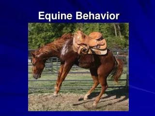
Equine Anatomy. Anatomy Terminology. The hip area is also called the hindquarter The topline is the back and loin from the wither to the croup (rump). The topline is also referred to as the length of the back. The underline is the area from elbow to stifle.

Equine Anatomy
E N D
Presentation Transcript
Anatomy Terminology • The hip area is also called the hindquarter • The topline is the back and loin from the wither to the croup (rump). The topline is also referred to as the length of the back. • The underline is the area from elbow to stifle
Skeletal and Muscular Information • Horses have approximately 210 bones • The horse’s skeleton has the mobility of its part to allow the horse much freedom of movement • The bones support muscles and protect internal organs.
Muscles are attached to bones by tendons and move the bones by contracting and relaxing • Tendons are encased in thin, fibrous sheets (tendon sheaths).
Parts of the Skeletal/Muscular System • Vertebral column- consists of bones in the tail, neck, back, loin and croup. • Ribs- 18 pairs in a horse, but 19 are frequently found on one side or the other. • Sternum- Canoe shaped breastbone made up of seven fused segments.
Skull- encloses the brain and important sense organs. • Thoracic limbs- all the bones of the front legs • Pelvic limbs- bones of the hind legs • Neck muscles- should be long, smooth and flat
Forearm muscles- should be long, lean and attach to the bone close to the knee. • Muscling in the horse’s back and loin add support in the vertebral column • Long, tapered muscles in the hindquarter provide speed whereas large, bulging muscles provide more power.
Functions of the skeletal system • The ribs provide protection and space for vital internal organs and should be long and well sprung. • The skull protects and provides cavities for the eyes and nervous system.
Thoracic limbs carry about 60% of the horse’s weight and are subject to a lot of stress and affect the movement, usefulness and value of the horse. • The pelvic limbs are the horse’s main means of propelling itself forward. • The stifle joint has a locking mechanism that allows the animal to sleep when standing • When the stifle joint sticks, the horse cannot bend the joint and the “stifled” horse requires surgery.
The hock joint is the most complex and most important single joint in the horse. • There are more serious unsoundness found in the hocks than any other part of the body • A horse must be structurally sound to move and perform well.
Functions of the muscular system • The neck muscle affects the ease and freedom of movement of the forelegs and should be long, smooth and flat. • Long, lean forearm muscles allow long strides. • Good muscling in the back and loin helps support the vertebral column and prevents “swayback” or a sagging back when the horse is used for riding.
Organs of the Thoracic Cavity • The thoracic cavity is the area between the neck and abdomen • Organs of the thoracic cavity include the circulatory and respiratory systems • Ribs- form the side of the thoracic cavity.
Heart- lies toward the bottom of the thoracic cavity and to the left of center. • Lungs- lie to the sides and behind the heart and fill most of the thoracic cavity.
Functions of the Thoracic Cavity • Heart pumps blood throughout the horse’s body. • The lungs exchange oxygen and carbon dioxide between the blood and the air.
Organs of the Abdominal Cavity • The abdominal cavity extends from just behind the thoracic cavity to the pelvic region.
Diaphragm- body partition of muscle and connective tissue. Separates the abdominal and thoracic cavities.
Major organs include • Liver- large organ extending all the way across the abdominal cavity. • Spleen and stomach- lie behind the liver and in front of the small and large intestines. • Kidneys- lie on each side of the backbone and under the last ribs in the loin area of the horse.
Functions of the Abdominal Cavity • The liver metabolizes several nutrients, detoxifies (removes the poison) many drugs and poisons and stores some nutrients. • The spleen and stomach are part of the gastrointestinal tract and function in the digestion and utilization of food. • The kidneys remove wastes from the body and conserve fluids and important components of the blood.
Organs of the Pelvic Cavity • The pelvic cavity is continuous with the abdominal cavity. • The rectum is the terminal portion of the intestine, which continues from the abdominal cavity to the pelvic cavity. • The urinary bladder lies within the pelvic cavity and extends into the abdominal cavity when full.
Major pelvic organs include: • Male reproductive organs- located toward the back and at the base of the pelvic cavity. or 2. Female reproductive organs – extend from the back of the cavity to near the abdominal cavity.
Functions of the Pelvic Cavity • The rectum is the terminal portion of the intestine and serves as the exit part for solid waste. • The urinary bladder collects liquid waste. • Reproduction
Digestive System Basics A. The digestive system of a horse is unique in that: • It has a relatively small but efficient stomach for grain utilization. • It has a large cecum and colon for roughage utilization.
B. The digestive system or alimentary canal of a horse: • Consists of a muscular hollow tube over 100 feet long that loops itself many times as it runs from the mouth to the anus. • The alimentary canal varies in size from 1 inch in diameter at the small intestine to 8 inches in diameter at the large colon.
Digestion is the process of breaking foodstuffs into their component nutrients so that they will be mostly soluble in water and easily absorbed (taken in) through the mucous membrane that lines the intestinal tract. There are several aspects of the process: 1. Mechanical action includes mastication (chewing), deglutition (swallowing), intestinal movements and defecation (elimination of waste).
2. Secretory action is the action on foodstuffs by secretions from glands within the body. 3. Chemical action would include action upon foodstuffs by the chemicals found in the stomach. 4. Microbial action is the breakdown of foodstuffs by very minute organisms within the digestive tract.
Digestion occurs from the time food is eschewed (ingested) until it is passed into the small colon for elimination from the rectum.
Anatomy of the Digestive System A. The mouth includes the teeth, the tongue and large salivary glands. • A mature horse secretes about 10 gallons of saliva a day. • Saliva wets foodstuff so that it is easier to go down the esophagus, and begins the digestion of carbohydrates.
B. The Pharynx is the muscular, funnel shaped upper back portion of the horse’s mouth designed to guide food down the esophagus.
C. The esophagus is a highly muscular 4-5 foot tube, which carries food and water from the mouth to the stomach through progressive waves of muscular contractions. • A horse cannot belch to relieve gas pressure of vomit as a result of the esophagus. • A horse’s stomach will usually rupture before vomiting occurs.
The stomach is a relatively small u-shaped sac at the front of the abdominal cavity. • Very little nutrient absorption and very little bacterial action occur in the stomach. • Swallowed foods are acted on by gastric juices secreted from glands in the mucous membrane lining the stomach to break down protein and fat. • Because of the small size of the stomach, horses need to be fed small amounts 2-3 times daily for efficient digestion.
The small intestine is a 2-inch by 60 foot tube that coils and loops its way from the stomach to the large intestine. • The small intestine along with the pancreas and liver supplies most of the enzymes for digestion. • The small intestine digests and absorbs 60-70% of the protein a horse eats and digests most of the soluble carbohydrates for the horse to use for energy. • The contents of the small intestine are about 92-95% water.
The Large Intestine is a 25 foot long tube comprised of the cecum, large colon, small colon and rectum, which moves undigested material from the small intestine to the anus for elimination. • Bacteria action is considerable in the large intestine to digest cellulose, produce amino acids for protein synthesis, and produce fatty acids which supply about one fourth of the horse’s energy. • The cecum is the primary site of water absorption.
The Rectum consists of the small colon and anus and receives feces that the small colon has formed. • Feces are formed in characteristic balls. • A horse on a standard diet of grain and hay voids 40-50 pounds of feces over the course of 8-12 times during the course of a day.
Digestion Progression • The stomach makes up only 10% of the total capacity of the digestive tract. • Food passes through the stomach in about 45 minutes • The stomach holds only 2-4 gallons.
The small intestine makes up 30% of the digestive tract capacity. • Food passage takes from 2.5 to 4 hours. • The small intestine holds about 12 gallons.
The large intestine makes up 50-60% of the total capacity of the digestive tract. • Food passage takes from 36 to 48 hours. • The combined capacity of the components of the large intestine is 30-35 gallons.