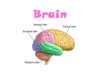
BRAIN
BRAIN. PHAKOMATOSIS. PHAKOMATOSIS. STURGE-WEBER SYNDROME VON HIPPEL-LINDAU DISEASE. STURGE-WEBER SYNDROME. Description:.


BRAIN
E N D
Presentation Transcript
BRAIN PHAKOMATOSIS
PHAKOMATOSIS • STURGE-WEBER SYNDROME • VON HIPPEL-LINDAU DISEASE
Description: • Sturge-Weber syndrome (encephalotrigeminal angiomatosis) is a congenital disorder characterized by localized atrophy and calcification of the cerebral cortex with an ipsilateral port wine-colored facial nevus in the area of the trigeminal nerve distribution.
Etiology : • Hereditary disorder attributed to autosomal dominant and autosomal recessive patterns.
Epidemiology: • Incidence is 1 per 1,000 patients in mental institutions.
Signs and Symptoms: • Patients may present with port-wine stain, seizure disorder, hemiatrophy, hemianopsia, mental retardation, and glaucoma.
Imaging Characteristics : CT • Cerebral atrophy may be seen. • Calcified areas of the brain will appear hyperdense. MRI • Cerebral atrophy is best seen on T1-weighted images. • Lower (hypointense) signal may be present in calcified areas of the brain. • FLAIR images are used to demonstrate leptomeningeal (pia mater and arachnoid) abnormality as a hyperintense signal.
Treatment: • Symptomatic treatment for the above-mentioned conditions.
Prognosis : • Most cases are considered to be mild and life expectancy is usually normal.
Figure 1. Sturge-Weber Syndrome. A B Noncontrast CT images of the brain (A) and bone window (B) demonstrate bilateral frontal and parietal cortical calcifications.
Figure 2. Sturge-Weber Syndrome. Postcontrast T1W axial (A) and coronal (B) images show cerebral atrophy.
VON HIPPEL-LINDAU DISEASE
Description: • An autosomal dominant (hereditary) condition characterized by angiomas of the retina and cerebellum, visceral cysts and malignancies, seizures and mental retardation.
Etiology : • Hereditary disorder.
Epidemiology: • Prevalence is estimated at 1 to 35,000 to 40,000 of the population. There is no predilection toward males or females.
Signs and Symptoms: • Patients typically become symptomatic in their third or fourth decade of life. Characterized by retinal angiomas, hemangioblastomas of the cerebellum and spinal cord, cystic disease of the kidneys, adrenal glands, or pancreas. Patients may experience seizures and mental retardation.
Imaging Characteristics : MRI • Tumors within the CNS appear as isointense to hypointense on T1-weighted images. • T2-weighted images present the tumors as hyperintense. • T1-weighted images with contrast demonstrate the tumor as hyperintense (the most sensitive means of detecting the CNS tumor).
Treatment: • Symptomatic treatment for the above-mentioned conditions and surgical intervention of tumors where and when appropriate.
Prognosis : • Varies depending on degree of symptoms and if cancer is detected. There is no known cure for this hereditary disease. Death is usually associated with complications of brain tumors or renal cancer.
Figure 1. Von Hippel-Lindau Disease. A B Axial T1W (A) shows a small hypointense nodule in the medial aspect of the left cerebellar hemisphere which enhanced with contrast administration T1W with gadolinium (B).
Figure 2. Von Hippel-Lindau Disease. A small enhancing nodule is seen on a sagittal T1W image with gadolinium. These are likely hemangioblastomas in this patient with Von Hippel-Lindau (VHL) Disease.
BRAIN VASCULAR DISEASE
VASCULAR DISEASE: • Arteriovenous Malformation • Intracranial Aneurysm • Intracerebral Hemorrhage (Hemorrhagic Stroke) • Ischemic Stroke (Cerebrovascular Accident) • Superior Sagittal Sinus Thrombosis
Description: • An arteriovenous malformation (AVM) is the most common type of vascular malformation and is characterized by direct artery-to-vein communication without an intervening capillary bed.
Etiology : • An AVM is a congenital lesion, which is the result of abnormal fetal development at approximately 3 weeks’ gestation.
Epidemiology: • Males generally present during middle age and are slightly more affected than females. Between 80% and 90% are located in the cerebrum, 10% and 20% located in the posterior fossa.
Signs and Symptoms: • Clinical presentation depends on the location and size of the AVM with most present between the second and third decade of life. By the age 50 years, 80% to 90% are located symptomatic. Hemorrhage will be present in about 50% of the cases. Other symptoms include seizures and headaches.
Imaging Characteristics : • Appears as a collection of “worms”. CT • Isodense to slightly hyperdense without contrast enhancement. • Calcification in 25% to 30%. • Atrophy. • Hyperdense serpentine appearing vessels with contrast enhancement. MRI • T1- and T2-weighted images demonstrate serpentine-appearing vessels with signal variations (flow void) in the vessels.
Treatment: • Depends on the age and general health of the patient. Endovascular embolization therapy, surgery intervention, stereotactic radiotherapy, or a combination of the above is useful in treating an AVM.
Prognosis : • The mortality rate is approximately 10% when hemorrhage is present.
Figure 1. Arteriovenous Malformation. A B Axial T1W Pre- (A) and postcontrast (B) enhanced images show a left frontal lobe mass with surrounding edema, minimal peripheral enhancement, and “worm”-like flow voids.
Figure 2. Arteriovenous Malformation. T2W axial image shows hyperintense edema around the mass “worm”-Ike flow voids.
Description: • An intracranial aneurysm is a localized dilation of a cerebral artery. The mostcommon form is the berry aneurysm, a saclike outpouching usually arising from at an arterial junction in the circle of Willis. Cerebral aneurysms often rupture and result in subarachnoid hemorrhage.
Etiology : • Weakening of the arterial wall may result from hemodynamic stresses. As an example, hypertension and atherosclerosis may restrict blood flow thus increasing blood pressure against an arterial wall, stretching it like an overblown balloon and making it likely to rupture. There is an increased incidence with: polycystic kidney disease, aortic coarctation, and family history.
Epidemiology: • Incidence rate is slightly higher in women than men. The peak age of occurrence is between 40 and 60 years. Anterior circulation is affected 90% of the time, while the vertebrobasilar circulation is affected only 10%.
Signs and Symptoms: • Intracranial aneurysms may go undetectable until they rupture; however, a very large nonruptured aneurysm can mimic the signs and symptoms of a tumor. If eh aneurysm ruptures, they usually present as a subarachnoid hemorrhage. Signs and symptoms may vary depending on the location and severity of the ruptured aneurysm. Other common signs and symptoms may include headaches, nausea and vomiting, hemiparesis or motor deficit, nuchal rigidity, loss of consciousness, and coma.
Imaging Characteristics : • Conventional angiography is the gold standard for the diagnosis of aneurysms. CT • In patients with ruptured intracranial aneurysm, a noncontrast study demonstrates a subarachnoid hemorrhage in the basilar cisterns as hyperdense in approximately 95% of the cases. • Contrast-enhanced CT may show very large aneurysms.
MRI • T1- and T2-weighted images appear with variable intensities (flow void). • Magnetic resonance angiography (MRA) can diagnose most large aneurysms (>5 mm).
Treatment: • Surgical intervention is best accomplished by a small metal clip or ligation around the neck of the aneurysm. Neuroradiologic intervention techniques also available for treatment of intracranial aneurysms include Guglielmi detachable (GD) coils.
Prognosis : • In event that the aneurysm ruptures, the prognosis may be determined by the severity of the initial hemorrhage, rebleeding of the aneurysm, vasospasm.
Figure 1. Intracranial Aneurysm. Computed tomographic angiography (CTA) 3D reconstruction shows a large right M1 middle cerebral artery (MCA) aneurysm.
Figure 2. Intracranial Aneurysm. A B Axial T1W pre- (A) and postcontrast (B) enhanced images show an enhancing mass in the region of the right MCA.
INTRACEREBRAL HEMORRHAGE (HEMORRHAGIC STROKE)
Description: • Intracerebral hemorrhage (ICH) occur when blood escapes from a ruptured vessel in the brain.
Etiology : • Results from a rupturing of a blood vessel, usually an artery, within the brain. Hemorrhagic infarcts are frequently associated with hypertension, arteriosclerosis, or an aneurysm. Other factors may include trauma, neoplasms (primary or metastasis), or drug use such as cocaine, amphetamine, and phenylpropanolamine.
Epidemiology: • Approximately 20% of all strokes are hemorrhagic.
Signs and Symptoms: • Patient may present with paralysis, motor weakness, headaches, or loss of consciousness.