Microprocessor-based Design for Biomedical Applications
This lecture discusses the origin and characteristics of bioelectric signals, electrodes and sensors, programming, and the physiological basis for action potentials. It also covers topics such as nerve cells, signal pathways in the central nervous system, and various bioelectric signal measurements including ECG, EMG, EOG, and EEG.

Microprocessor-based Design for Biomedical Applications
E N D
Presentation Transcript
Microprocessor based Design for Biomedical ApplicationsMBE 3 – MDBAV : Bioelectric SignalsCharacteristics
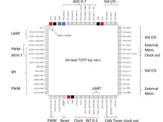
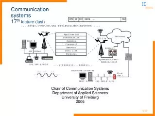
last Lecture:Interrupt driven Uart Communication Setup stdio- functions, printf Atmega8 Analog Digital Conveter Analog Comparator
Today: Origin and characteristics of bioelectric signals Electrodes and sensors Review of Project exercises Programming
Electrical and chemical gradients at the semi-permeable cell membrane
Electrical and chemical gradients at the semi-permeable cell membrane
Nernst – equation (chemical potential): R … Gas-Constant = 8,3143 J / (mol·K) T … Temperature (Kelvin) Goldman – equation (for different ions): As a result, we get a membrane resting potential of about -70mV
Depolarization Sodium Cations rush in Hyperpolarization Potassom Cations rush out
Maintaining the Resting potential Sodium/potassum Ion pump
Voltage- and Time dependent activation of Ion Channels: the physiological basis for action potentials Sodium-Channel Potassum- Channel
Hodgkin - Huxley Model (1952) ●Researched the Giant Squid-Axon ●Used the Voltage-Clamp technique -> Isolation of channel currents of Na und K ●Developed a model for the function of the channel proteines Alan Hodgkin Andrew Huxley
Sodium and Potassum conductance Calculated by the Hodkin Huxley Model (curve), measured (dots)
Action potential: the result of Na and K Ion movement through the membrane
Axo-dendritic transmission of action potentials, Synaptic transduction
Signal Pathways in the central nervous system
ECG Electro-Cardiogram, Heart activityEMG Electro-Myogram, Muscle movementEOG Electro-Oculogram, Eye movementEEG Electro-EncephalogramGSRGalvanic Skin Response ● Measured with electrodes: skin-electrode interface: Ions <--> Electrodes Breathing, temperature, movement etc. ● Measured with other sensors / transducers: NTC, LDR, piezo-crystal, hall-sensor, Accelerometer, Goniometer, …
ECG - applications ● Diagnostics ●Functional analysis ●Implants (pace maker) ●Biofeedback (Heartrate variability, HRV) ● Peak Performacne Training, Monitoring
EMG surface (glue-) electrodes EMG - signal (up to 3mV, 1kHz)
EMG electrodes (active) EMG electrodes (passive)
EMG electrodes (active)
Needle electrodes adhesive electrode
EMG - applications ● Rehabilitation ●Functional analysis ●active Prothetics, Orthesis ●Biomechanics, Sports medicine
EOG - applications ● Diagnostics ●Functional analysis ●Human Computer Interfaces
EEG Electrode – cap locations of the 10/20 system
Unipolar measurement( indifferential right ear electrode ) Bipolar measurement
EEG, Alpha bursts when eyes closed, alpha desynchronisation when eyes opened
Quantitative EEG (QEEG),many EEG channels (up to 256)source / dipole localisation
Auditory Evoked Potentials (AEP) Trial averaging Also: VEP SSEP