
edema-
E N D
Presentation Transcript
Edema is not accompanied with cellular edema Isotonic fluid Ⅰ. concept • Edema: is the accumulation of excessive body fluid in interstitial space or serous body cavity, which is a pathologic process caused by diseases rather than a disease entity. • Hydrops: when body fluid is accumulated in serous body cavity, edema is also called hydrops, such as ascites and hydrothorax.
Ⅱ. classification • According to the range that edema fluid spreads to: • Anasarca (generalized edema) • Local edema • According to the cause of edema: • Renal edema • Hepatic edema • Cardiac edema • Malnutritional edema • lymphedema
Edema: is the accumulation of excessive body fluid in interstitial space or serous body cavity intake output Kidney(urine) 1500ml Intestine(feces) 100ml Lung(water vapor) 400mlSkin(sweat ) 500ml Ⅲ. Etiology and pathogenesis Im • balance of fluid exchange between plasma and interstitial compartment Im • balance of fluid exchange between extra- and intra-body Renal retention of sodium and water Urine volume ICF 吸收水分 Interstitial fluid 40%BW Plasma 15%BW 5%BW
(Ⅰ). imbalance of fluid exchange between plasma and interstitial compartment
capillaries When blood flows through capillary, fluid exchange, including water and some small molecules that can pass through the capillary wall, will happen between plasma and interstitial compartment.
Total Pressure Differences Inside and Outside Capillary obstruction ↑ ↑ ↓ permeability The force promoting fluid outshift from capillary to interstitial compartment includes blood pressure and negative interstitial pressure. The force attracting fluid from interstitial compartment to capillary is blood colloid osmotic pressue. At the arterial end, the sum of the forces causes fluid to move from the capillary into the tissue. At the venous end, the sum of the forces causes fluid to move into the capillary. About nine-tenths of the fluid that leaves the capillary at its arterial end reenters the capillary at its venous end. About one-tenth of fluid passes into the lymphatics. Figure 7-7
↑capillary blood pressure ↑force driving fluid into interstitium ↑formation of interstitial fluid When greater than lymphatic compensatory return edema 1. Increased capillary blood pressure • Causes: • Elevated plasma volume • Increased venous pressure • - Increased general venous pressure, i.e. congestive heart failure • - Increased local venous pressure, i.e. venous thrombosis • Arteriolar dilation i.e. acute imflammation
2. Decreased plasma colloid osmotic pressure ↓ plasma colloid osmotic pressure • Causes: plasma albumin content decrease • Decrease of protein production i.e. hepatic cirrhosis, malnutrition • Excessive loss of protein i.e. nephrosis • Elevated catabolism of protein i.e. chronic debilitating diseases, such as malignant tumor ↓force drawing water back into capillary from interstitium ↑formation of interstitial fluid When greater than lymphatic compensatory return edema
3. Obstruction of lymphtic During the process of interstitial fluid formation, one-tenth of fluid that leaves the capillary at its arterial end returns to the venous circulation via lymphatics. So obstruction of lymphatic will result in edema. • Causes: • Blockage by cancer • Blockage by infection, especially with filarial
4. Increased capillary permeability ↑capillary permeability • Causes: inflammation • Infection • Burn • Allergic response • Trauma • Anoxia • Acidosis Filtration of more protein from capillary to interstitium ↓Plasma colloid osmotic pressure ↑formation of interstitial fluid When greater than lymphatic compensatory return edema
(Ⅱ). Imbalance of fluid exchange between extra- and intra-body------ renal retention of sodium and water
Glomerular( filtration) and tubular (reabsorption) balance (G-T balance) • In normal condition, 99-99.5% of total volume of sodium and water filtrated via glomeruli are reabsorbed by tubules. • 60-70% of filtrates are actively reabsorbed by proximal convoluted tubule. • The reabsorptions of sodium and water at distal tubule and collectiong duct are regulated by hormone.
GFR is decreased, while tubular reabsorption is not decreased accordingly; • Tubular reabsorption increases, while GRF isn’t increased. G-T imbalance Retention of sodium and water • ↓GFR • ↑Reabsorption of proximal tubule • ↑Reabsorption of distal tubule and collection tubule
1. Decreased glomerular filtration rate (GFR) • When decreased GFR is not accompanied with decreased tubular reabsorption, retention of sodium and water will be caused.
Factors determining the GFR: • Filtration area and membrane permeability • Filtration pressure • Effective circulating blood volume or renal blood volume
Renin- angiotensin system Sympathetic - adrenal medullary system ↓Renal blood volume ↓GFR 1. ↓GFR Causes • Extensive glomerular damage i.e. Acute or chronic glomerulonephritis • Decrease of effective circulating blood volume i.e. congestive heart failure, nephrotic syndrome
glomerular filtration rate (GFR) FF = renal plasma flow (RPF) 2. -Increased reabsorption in proximal tubule Increased filtration fraction (FF) GFR: amount of plasma filtered at glomerulus into Bowman’s capsule FF is the fraction of renal plasma flow that is filtered at the glomerulus In normal condition: FF: 20%
Increased FF make elevated reabsorption of proximal tubule Increased FF the protein concentration in the plasma entering the peritubular capillaries increases The peritubular capillary oncotic pressure increases enhancing fluid reabsorption from the renal interstitial space to the capillary decreases renal interstitial pressure favoring reabsorption across the tubular epithelium and minimizing back flux from the renal interstitial space to the tubule lumen. ↑Reabsorption in proximal tubule
Causes of FF increasing Congestive heart failure Nephrotic syndrome Decreased effective circulatory blood volume Sympathetic-adrenal medullary system exciting Efferent arteriole constricts stronger than afferent one ↑Efferent arteriole resistance GFR is increased relative to renal plasma flow ↑FF
Congestive heart failure Nephrotic syndrome ↓Stimulation of volume-receptor in left atrim and thoracic vessel ↑ADH secretion Retention of sodium and water ↓Effective circulatory blood volume Renin-angiotensin-aldosterone systm activation Sympathetic nerve excitation ↑Secretion of renin by Juxtaglomerular cell ↓Renal perfusion pressure ↓Renal blood flow ↓GFR→sodium at macula densa increased ADH and ADS secretion 3. -Increased reabsorption in distal tubule and collecting duct
Renin Adrenal gland Ang Ⅰ Ang Ⅱ The renin-angiotensin-aldosterone system
question • Why can congestive heart failure make edema? • ↑General venous pressure • ↓Plasma colloid osmotic pressur because of dilution of blood • Dysfunction of lymphatic return because of increased venous pressure • ↓GFR • ↑FF • ↑ADH and ↑ADS
1. The feature of edema fluid • Transudates • Formed under almost normal capillary permeability • Low content of cells and protein • Exception: edema fluid due to lymphatic obstruction • Exudates • Resulted from an increase in capillary permeability • Contain plentifu cells and a large amount of protein • Usually seen in inflammation
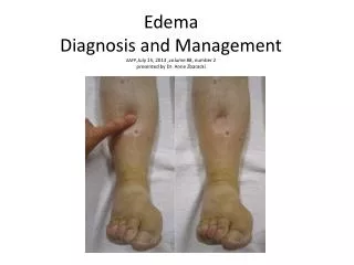
2. The cutaneous characters of edema • Pitting edema • a tissue area is pressed by thumb and after the thumb is removed, a pit is left at the pressed site for a few seconds. • Recessive edema • Edema without pit. • Before pitting edema emergeing, excessive interstitial fluids already exist in generalized edema patient. Increased extra interstitial fluid is bound in a proteoglycan filament messwork of tissue, so that this gel fluid does not flow easily through the tissues.
3. Distribution character of generalize edema Influencing factors • Gravity • Structure of tissue • Local hemodynamics • Cardiac edema: ankle • Renal edema: eyelids and face • Hepatic edema: ascites
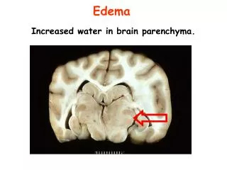
4. Effect of edema • Generally, edema is harmfu, i.e. • Impedes nutritional supply to cells • Edema of brain is life-threatening, intracranial pressure increase, herniation • Protect effect: the edeme fluid of inflammatory edema can dilute, neutralize toxin and transport the antibody.