Download

1 / 35
350 likes | 577 Views
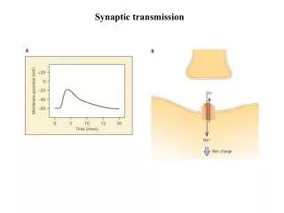
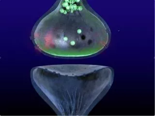
A readily retrievable pool of synaptic vesicles. GIRARDEAU Paul SILVESTRE DE FERRON Benoit. Synaptic transmission. Introduction. Synaptic transmission is limited by the r ecycling of synaptic vesicles for many rounds of use. Synaptic transmission. Introduction.

E N D
A readilyretrievable pool of synapticvesicles GIRARDEAU Paul SILVESTRE DE FERRON Benoit
Synaptic transmission Introduction Synaptic transmission islimited by the recycling of synapticvesicles for many rounds of use
Synaptic transmission Introduction The main process of vesiclerecyclingismediated by clathrin The formation of clathrin-coated vesicles from the plasma membrane BUT slow process (sec to mn)
Vesiclerecycling Introduction ContinuousactivityNecessity of rapidrecycling 2 mechanismsidentified Clathrin-independent: Kiss and run Clathrinmediated: Preassembled structures Pool of preassembledvesicleproteins At the pre-synaptic surface What are the temporal dynamics of such a « readilyretrievable pool » ?
Vesicleproteins Introduction VARIOUS SYNAPTIC PROTEINS AT THE SYNAPTIC VESICULAR MEMBRANE Synatptotagmin (Syt1) and Synaptobrevin (Syb2) : mostnumerousvesicularproteins VGAT (vesicular transporter of aminoacids in inhibitoryneurons) • Labeling: • of Syt1, Syb 2 and VGAT • to test the hypothesis of the pool of preassembled structures • in rat hippocampal neurons
Exo-endoxytosis of endogenoussynapticvesicleproteins Methods Synaptotagmin 1 antibodycoupledwithcypHer (pH sensitive fluorophore) anti-Syt1-cypHer Cypheremitsred fluorescence whenexcitedat 640 nm The fluorescence is maximal at pH 5.5 (pH of insidesynapticvesicle) Normalized fluorescence of anti-Syt1-cypHer VGAT antibodycoupledwithcypHer (pH sensitive fluorophore) anti-VGAT-cypHer For studyinginhibitory synapses
Exo-endoxytosis of endogenoussynapticvesicleproteins Results Spontaneousactivity pH 7,4 7,3 7,2 7,1 7,0 6,9 6,8 6,7 6,6 6,5 6,4 6,3 6,2 6,1 6,0 5,9 5,8 5,7 5,6 5,5 Synaptotagmin 1 antibodycoupledwithcypHer (pH sensitive fluorophore) Synaptotagmin 1 Red fluorescence emission
Exo-endoxytosis of endogenoussynapticvesicleproteins Results pH 5.5 VGAT antibodycoupledwithcypHer (pH sensitive fluorophore) VGAT Red fluorescence emission
Exo-endoxytosis of endogenoussynapticvesicleproteins Results Fluorescence image of hippocampalneuronslabeledwith anti-VGAT-cypHer
Exo-endoxytosis of endogenoussynapticvesicleproteins Question Are these probes efficiency to report stimulation-dependent exo-endocytosis?
7,4 7,3 7,2 7,1 7,0 6,9 6,8 6,7 6,6 6,5 6,4 6,3 6,2 6,1 6,0 5,9 5,8 5,7 5,6 5,5 Exo-endoxytosis of endogenoussynapticvesicleproteins Results ElicitedAPsat 20 Hz (50, 200, 600, 900) Synaptotagmin 1 antibodycoupledwithcypHer (pH sensitive fluorophore) -> Anti-Syt1-cypHer pH Synaptotagmin 1 Red fluorescence emission
Exo-endoxytosis of endogenoussynapticvesicleproteins Results ElicitedAPsat 20 Hz (50, 200, 600, 900) Synaptotagmin 1 antibodycoupledwithcypHer (pH sensitive fluorophore) -> Anti-Syt1-cypHer Synaptotagmin 1 Red fluorescence emission pH 7,4 7,3 7,2 7,1 7,0 6,9 6,8 6,7 6,6 6,5 6,4 6,3 6,2 6,1 6,0 5,9 5,8 5,7 5,6 5,5
Exo-endoxytosis of endogenoussynapticvesicleproteins Results These probes are efficiency to report stimulation dependent exo-endocytosis
Size of the surface pool of synapticvesicleconstituents Results 50 APs 100 APs 200 APs Previousstudies : Endocytic rate after stimulus = Endocytic rate during the stimulus 200 APs Fluorescence variations riselinearlywith stimulus strength
Size of the surface pool of synapticvesicleconstituents Question Are endogenoussynapticvesicleproteinsalreadypresent on the presynaptic membrane?
Size of the surface pool of synapticvesicleconstituents Results Synaptotagmin 1 antibodycoupledwithcypHer (pH sensitive fluorophore) -> Anti-Syt1-cypHer pH Synaptotagmin 1 7,4 7,3 7,2 7,1 7,0 6,9 6,8 6,7 6,6 6,5 6,4 6,3 6,2 6,1 6,0 5,9 5,8 5,7 5,6 5,5 Red fluorescence emission Buffer of pH 5,5 Buffer of pH 5,5 Proteinsalreadypresents on the presynaptic membrane Number of boutons ΔF = size of the surface pool ΔF ΔF ~ 50 a.u. Theseproteins are able of compensatingexocytosisinduced by 70 APs
Labeledantibodies report samerecyclingkinectics as spH Methods Dual-colorimaging • Exogenous probe Synaptotagmin 1 antibodycoupledwithcypHer (pH sensitive fluorophore) anti-Syt1-cypHer Fluorescence in acidiccompartments • Overexpressed probe Synaptobrevin 2 coupledwithpHluorin (pH sensitive GFP) SpH Fluorescence in neutralcompartments
Labeledantibodies report samerecyclingkinectics as spH Results 200 APsat 20 Hz Synaptotagmin 1 antibodycoupledwithcypHer (pH sensitive fluorophore) -> Anti-Syt1-cypHer pH 5,5 Synaptotagmin 1 Red fluorescence emission Synaptobrevin 2 coupledwithpHluorin(pH sensitive fluorophore) -> SpH Green fluorescence emission 7,4
Labeledantibodies report samerecyclingkinectics as spH Results 200 APsat 20 Hz Synaptotagmin 1 antibodycoupledwithcypHer (pH sensitive fluorophore) -> Anti-Syt1-cypHer 5,5 Synaptotagmin 1 Red fluorescence emission Synaptobrevin 2 coupledwithpHluorin(pH sensitive fluorophore) -> SpH pH Green fluorescence emission 7,4 Mirror-image signals These probes report exo-endocytosis
Labeledantibodies report samerecyclingkinectics as spH Results Sameresults in inhibitory synapses with VGAT
A surface RRetP of synapticvesicleconstituents Question Vesicleproteinsalreadypresent on the presynaptic membrane Are synapticvesicleproteins, exo- and endocytosed by the same stimulus, identical or different?
A surface RRetP of synapticvesicleconstituents Methods • First experiment Inactivation of Synaptobrevin 2 fluorescence on the presynaptic membrane TEV cleavage site betweenSynaptobrevin 2 and pHluorin • Second experiment Inactivation of Synaptotagmin 1 fluorescence on the vesicles Photobleaching
A surface RRetP of synapticvesicleconstituents Results 50 APsat 20 Hz Synaptotagmin 1 antibodycoupledwithcypHer (pH sensitive fluorophore) -> Anti-Syt1-cypHer pH 5,5 Synaptotagmin 1 Synaptobrevin 2 coupledwithpHluorin (pH sensitive fluorophore) -> SpH TEV cleavage site pH Tobacco etch virus (TEV) protease 7,4 A surface pool of vesicleproteinsisendocytosed
A surface RRetP of synapticvesicleconstituents Results 50 APsat 20 Hz Photobleaching pH 5,5 Synaptotagmin 1 antibodycoupledwith cypHer (pH sensitive fluorophore) -> Anti-Syt1-cypHer Synaptotagmin 1 pH Synaptobrevin 2 coupledwithpHluorin (pH sensitive fluorophore) -> SpH 7,4 Presortedsynapticvesicleproteins are preferentiallyendocytosed on exocytosis
A surface RRetP of synapticvesicleconstituents Results WithspHoverexpression WithoutspHoverexpression Endocytosis of Syt1 or VGAT is not perturbed by spHoverexression
A surface RRetP of synapticvesicleconstituents Results ReadilyReleasable Pool (RRP) Partial bleaching of the surface pool ReadilyRetrievable Pool (RRetP) RRP seems to becounterbalanced by an RRetP of similar size
Spatial organization of the RRetP Question How is the functionnal surface pool spatiallyorganizedat the presynapse?
Spatial organization of the RRetP Methods First labeling Second labeling Antibody to Synaptotagmin 1 Secondaryantibody Antibody to Synaptotagmin 1 Secondaryantibody Antibody to Homer1 (Post SynapticDensity) Secondaryantibody Antibody to RIM (Active Zone) Secondaryantibody IsoSTEDmicroscopy 4Pi microscopy
Spatial organization of the RRetP Results ? Presynaptic Postsynaptic R RIM (AZ) R H Homer1 (PSD) H R H R H Syt1 (?) IsoSTED
Spatial organization of the RRetP Results ? α Presynaptic Postsynaptic R RIM R H d R Homer1 H H R H Syt1 Axon Doghnut-like arrangement around the active zone of the RRetP
and conclusion Discussion Vesicleproteinsalreadypresents on the presynaptic membrane Presortedsynapticvesicleproteins are preferentiallyendocytosed on exocytosis RRP seems to becounterbalanced by an RRetP of similar size Doghnut-like arrangement around the active zone of the RRetP
and conclusion Discussion
A surface RRetP of synapticvesicleconstituents Results 50 APs 200 APs Incompletedepletion of the RRetPduring the 1st stimulus Partial replenishment of the RRetPafter the 1st stimulus