Download
1 / 28
280 likes | 500 Views
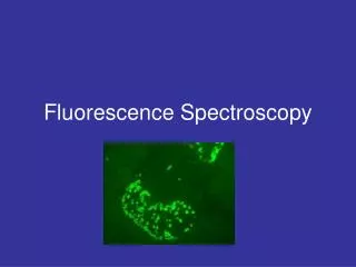
Explore the fundamental principles, applications, and technologies of fluorescence spectroscopy and microscopy for advancements in biology and medicine. Discover the delicate interactions at the molecular level and dynamic processes in biological systems.

E N D
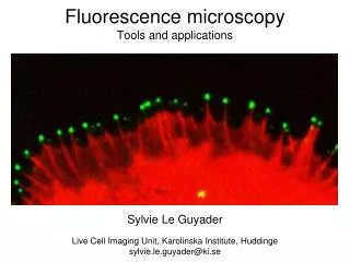
CZECH TECHNICAL UNIVERSITY IN PRAGUE FACULTY OF BIOMEDICAL ENGINEERING Fluorescence spectroscopy and microscopy for biology and medicine Martin Hof, Radek Macháň
CZECH TECHNICAL UNIVERSITY IN PRAGUE FACULTY OF BIOMEDICAL ENGINEERING Fluorescence spectroscopy and microscopy for biology and medicine Martin Hof, Radek Macháň Absorption of light and electronic transitions Basic principles of fluorescence, fluorescence spectra Lifetime of fluorescence and its measurement Quenching of fluorescence and its biological applications Anisotropy of fluorescence and its biological applications Influence of solvent on fluorescence spectra Foerster resonance energy transfer and excimer fluorescence Fluorescent proteins Fluorescence microscopy, confocal and 2-photon microscopy Resolution of fluorescence microscope and its enhancement Fluorescence correlation spectroscopy Photodynamic Therapy
Lakowicz J.R.: Principles of Fluorescence Spectroscopy, 3rd edn. Springer 2006cfs.umbi.umd.edu/ • Hof M., Hutterer R., Fidler V.: Fluorescence Spectroscopy in Biology. Springer Verlag • Gauglitz G., Vo-Dinh T.: Handbook of Spectroscopy. Wiley VCH Verlag, Weinheim 2003 • Prosser V. a kol.: Experimentální metody biofyziky. Academia,Praha 1989 • Invitrogen Tutorials www.invitrogen.com/site/us/en/home/support/Tutorials.html • Becker W.: The bh TCSPC Handbook http://www.becker-hickl.com/literature.htm Basic literature:
electric fields ions Fluorescent Probe viscosity temperature polarity pH Why fluorescence? Also fluorescence is very, very, very sensitive! Work with subnanomolar concentrations is routine while femtomolar and even SINGLE MOLECULE studies are possible with some effort • it provides information on the molecular environment • it provides information on dynamic processes on the nanosecond timescale Fluorescence Probes are essentially molecular stopwatches which monitor dynamic events which occur during the excited state lifetime – such as movements of proteins or protein domains
Experimental Systems Molecular structure and dynamics Cell organization and function Actin Mitochondria Nucleus Biological membrane Multicellular organisms GFP in a mouse
Instrumentation Microscopes Fluorimeters High throughput platereaders
1. Sir Isaac Newton1672: Showed that the component colors of the visible portion of white light can be separated through a prism, which acts to bend the light (refraction) in differing degrees according to the wavelength. Developed a “corpuscular” theory of light . 2. Christian Huygens 1692: Developed a wave theory of light A very brief history of the study of light 3. Hans Christian Oersted 1820 Showed that there is a magnetic field associated with the flow of electric current 4. Michael Faraday 1831 Showed the converse i.e. that there is an electric current associated with a change of magnetic field
5. James Clark Maxwell: 1865 Published his “Dynamical theory of the electromagnetic field” which combined thediscoveries of Newton, Young, Foucault, Oersted and Faraday into a unified theory of electromagnetic radiation Light consists of electromagnetic transverse waves of frequencyand wavelengthrelated by = ncwhere nis the index of refraction of the medium andcis thespeed of the light in vacuumc= 3x1010 cm/s B E we are interested in interactions of the electric field with the matter
6. Max Karl Ernst Ludwig Planck: 1900 Explained the laws of black body radiation by postulating that electromagnetic radiation is emitted at discrete energetic quanta E = hn , where Planck constant h =6.6256 *10-34 Js. 7. Albert Einstein: 1905 Explained the explained the photoelectric effect by assuming that light is adsorbed at discrete energetic quanta E = hn , photons. 8. Louis de Broglie: 1924 Introduced properties of electromagnetic waves to all particles – the wave-corpuscular dualism of quantum physics. A freely moving particle of momentum p has wavelength l=h/p.
Visible light X-ray Microwave UV IR Radio Wavelength Frequency Hz 1021 1013 1017 1011 1019 109 107 1015 Wavenumber cm-1 nm 1011 103 107 101 109 10-1 10-3 105 108 102 10-4 104 1010 10-2 100 106 Energy Kcal Energy eV 107 108 100 10-1 103 104 10-2 10-3 105 106 10-4 10-5 10-7 10-6 101 102 Wavelength and energy scale, appropriate units
Visible light UV IR nm 108 102 10-4 104 1010 10-2 100 106 The optical region of the electromagnetic spectrum Wavelength nm wavelength << optical elements molecules<< wavelength the whole molecules sense the same phase of light (vs. X-ray diffraction) vs. microwave or r.f. techniques
+ - Interaction of electromagnetic waves with matter • Atoms and molecules described as electric multipoles, first approximation: electric dipole • Classical electrodynamics: dipoles oscillate at the frequency of the external electromagnetic field Elastic scattering of light
Interaction of molecules with photons - quantum description • Light exists in form of discrete quanta –photons E = hn • Atoms and molecules occupy discrete energetic states, which can be found as the solution of Schroedinger’s equation. • Exchange of energy with photons is accompanied by transitions between those states. rotational states DJ = 1 microwave region vibrational states DN = 1 IR – VIS region E electronic states UV – VIS region
2 spontaneous emission induced emission absorption 1 Interaction of light with matter – overview of processes • inelastic (Raman) scattering – the photon either gives a part of its energy to the molecule or vice versa • elastic scattering – no exchange of energy between the molecule and the photon • absorption or emission of photons by the molecule • induced emission is coherent with incident light • spontaneous emission by individual molecules is incoherent • scattering is coherent and instantaneous
Elastic scattering of light • Rayleigh scattering – small molecules (x<0.3) as a “point dipole”, Isc ≈ n4 blue sky, red sunset x = 0.07 • Larger scatterers – macromolecules, cells, Mie theory for spherically symmetrical scatterers x = 7 http://omlc.ogi.edu/calc/mie_calc.html
Raman scattering • 1923 theoretically predicted by Adolf Smekal using classical physics • 1928 observed by C. V. Raman C.V. Raman (1888-1970) the photon and the molecule exchange energy n0-Dn n0+Dn n0 the photon is not absorbed: scattering is an instantaneous and coherent v2 hDn v1 elastic Stokes anti-Stokes branch of Raman spectrum
Raman spectrum intensity of Stokes branch is higher by a factor anti-Stokes Stokes
angle between polarization and D12, for random orientation of molecules Absorption of light S 2 Nf2 molecules DE = hv0 1 Nf1 molecules I dx I0 • electromagnetic energy density • F shape of the spectral line – conservation of energy • M – number of photons • small energy approximation – assumes that absorption does not change f2/f1=exp(DE/kT)=f(T) • N – number of molecules N = c NAS dx
Absorption of light S 2 Nf2 molecules DE = hv0 1 Nf1 molecules I dx I0 the Lambert Beer law e the molar extinction coefficient (molar absorptivity)
sample I PMT sample Deuterium/ Tungsten Lamp Mono- chromator Io PMT reference blank Absorption: measurement The Beer Lambert Law Absorption (Optical Density) = log Io / I = c l l is the path length of the sample (1 cm) • a typical sample: a solution in a cuvette • the solvent and the reflection from the cuvette walls contribute to the extinction of light • relative measurement of absorption Detector
Electronic transitions from the ground state to the excited state Energy S1 Probability HIGH HIGH MEDIUM LOW v1 3 v 12 v 11 v1 0 G Probability v 3 v 2 v 1 v 0 Inter-nuclear distance Wavelength nm
S1 v1 3 v 12 v 11 v1 0 G v 3 v 2 v 1 Inter-nuclear distance v 0 Electronic transitions from the ground state to the excited state Shaded areas reflects theprobability of where the electron would be if it were in that vibrational band Most favored transitions occur From the maximum shaded areas of the ground state To the maximum shadedareas of theexcited state
Electronic – vibrational spectrum other transitions (other vibrational modes, non-fundamental transitions,…) effect of room temperature effect of molecular surroundings
Increasing the number of double bonds shifts the absorption to lower energy N=5 5 pi-bonds, 10 electrons N=4 4 pi-bonds, 8 electrons molar absorbtivity N=3 3 pi-bonds, 6 electrons Wavelength nm Absorption maxima : The importance of conjugation The wavelength value of theabsorption maximum and themolar absorptivity are determined by the degree ofConjugatation of p-bonds
Benzene < Naphthalene < Anthracene < naphthacene < pentacene Abs. Max 262nm 275 nm 375 nm 475 nm 580 nm Log e 3.84 3.75 3.90 4.05 4.20 (Extinction) Log Extinction Coefficient 275 nm 375 nm 475nm absorption wavelength Increasing the number of aromatic rings increases the absorption maximum As the degree of conjugation increases (i.e the number of electrons involved in the delocalized p-orbitals) the absorption energy decreases(> l, the energy between the ground and excited state decreases) the absorption becomes more intense(>e, increased probability of absorption)
Emission of light - Luminescence Luminescence – the excess of light emitted above thermal radiation. The emission follows after the molecule has resided for some time in the excited state. according to excitation mechanism: photoluminescence – absorption of light chemiluminescence – chemical reaction thermoluminescence – heat electroluminescence – electric current … fluorescence phosphorescence photoluminescence – absorption of light
Typical sources of luminescence • organic molecules (usually with conjugatedp-bonds) – synthetic fluorophores (fluorescein, rhodamine, …), biological molecules (aromatic amino acids – Trp, Tyr, chlorophyll, …) • small inorganic molecules – noble gases (in discharge lamps), N2 (in lasers, responsible for bluish colour of spark discharges), … • inorganic crystals (diamond, Si, GaAs, … ) – the spectra depend on the bandgap size, which depends on the size of the crystal (nanocrystals emit in VIS – quantum dots), extreme photostability quantum dots – same material, different sizes
Acknowledgement The course was inspired by courses of: Prof. David M. Jameson, Ph.D. Prof. RNDr. Jaromír Plášek, Csc. Prof. William Reusch Financial support from the grant: FRVŠ 33/119970