Download
1 / 20
200 likes | 300 Views
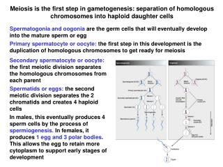
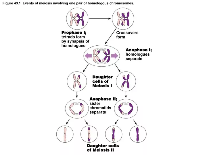
Prophase I; tetrads form by synapsis of homologues. Crossovers form. Anaphase I; homologues separate. Figure 43.1 Events of meiosis involving one pair of homologous chromosomes. Daughter cells of Meiosis I. Anaphase II; sister chromatids separate. Daughter cells of Meiosis II.

E N D
Prophase I; tetrads form by synapsis of homologues Crossovers form Anaphase I; homologues separate Figure 43.1 Events of meiosis involving one pair of homologous chromosomes. Daughter cells of Meiosis I Anaphase II; sister chromatids separate Daughter cells of Meiosis II
Figure 43.2a Spermatogenesis. Spermatogonium (2n) Mitosis Primary spermatocyte (2n) Growth phase Primary spermatocyte (2n) Meiosis I (DNA replicated before division) Secondary spermatocyte (n) Meiosis II (DNA not replicated before division) Spermatids (n) Spermiogenesis Functional sperm (n)
Figure 42.2 Structure of the testis. Immature sperm Interstitial endocrine cells Spermatic cord Blood vessels and nerves Seminiferous tubule Ductus (vas) deferens Head of epididymis Efferent ductule Spermatogenic cells Rete testis Lobule Straight tubule Septum Tunica albuginea Spermatic cord Body of epididymis Tunica vaginalis Cavity of tunica vaginalis Duct of epididymis Epididymis Tail of epididymis Testis
Figure 43.2b Spermatogenesis. Spermatogonia Primary spermatocytes Spermatids Immature sperm in lumen Sustentocytes (of testis)
Figure 42.5 Cross section of epididymis (120). Pseudostratified columnar epithelium Spermatozoa Stereocilia
Figure 42.3 Transverse section of the penis (3). Corpora cavernosa Venous cavities Tunic albuginea (surrounds corpora) Lumen of urethra Corpus spongiosum
Figure 43.3 Sperm in semen. Fluid medium of semen Tail Midpiece Sperm Head with acrosome
Figure 43.4 Oogenesis. Meiotic events Follicle development in ovary Oogonium (stem cell) Before birth 2n Follicle cells Mitosis Oocyte Primary oocyte 2n Primordial follicle Infancy and childhood (ovary functionally inactive) Primary oocyte (arrested in prophase I; present at birth) 2n Primordial follicle Each month from puberty to menopause Primary follicle Primary oocyte (still arrested in prophase I) 2n Secondary follicle Spindle Vesicular (antral) follicle Meiosis I (completed by one primary oocyte each month in response to LH surge) Secondary oocyte (arrested in metaphase II) n First polar body Ovulation Sperm Meiosis II of polar body (may or may not occur) Ovulated secondary oocyte Meiosis II completed (only if sperm penetration occurs) In absense of fertilization, ruptured follicle becomes a corpus luteum and ultimately degenerates. n n n n Polar bodies (all polar bodies degenerate) Second polar body Degenerating corpus luteum Ovum
Figure 43.5 Anatomy of the human ovary. Tunica albuginea Secondary follicle Late secondary follicle Granulosa cells Cortex Degenerating corpus luteum (corpus albicans) Mesovarium and blood vessels Germinal epithelium Vesicular (antral) follicle Primary follicles Antrum Secondary oocyte Ovarian ligament Zona pellucida Theca folliculi Medulla Ovulated secondary oocyte Corpus luteum Corona radiata Developing corpus luteum
Figure 42.9 Cross-sectional view of the uterine tube (12). Serosa Smooth muscle Highly folded mucosa Lumen
Figure 42.8 Cross-sectional view of the uterine wall. Endometrium Myometrium Serosa
Figure 43.6 Endometrial changes during the menstrual cycle. Necrotic (areas of dead and dying cells) fragments of functional layer of endometrium Elaborated glands Endometrium Glands Myometrium Endometrium • Functional layer • Basal layer Myometrium
Figure 43.7 Correlation of anterior pituitary and ovarian hormones with structural changes in the ovary and uterus. Fluctuation of gonadotropin levels: Fluctuating levels of pituitary gonadotropins (follicle-stimulating hormone and luteinizing hormone) in the blood regulate the events of the ovarian cycle. LH Plasma hormone level FSH Ovarian cycle: Structural changes in the ovarian follicles during the ovarian cycle are correlated with (d) changes in the endometrium of the uterus during the uterine cycle. Primary follicle Vesicular follicle Corpus luteum Secondary follicle Ovulation Degenerating corpus luteum Follicular phase Luteal phase Ovulation (Day 14) Fluctuation of ovarian hormone levels: Fluctuating levels of ovarian hormones (estrogens and progesterone) cause the endometrial changes of the uterine cycle. The high estrogen levels are also responsible for the LH/FSH surge in (a). Plasma hormone level Estrogens Progesterone The three phases of the uterine cycle: Endometrial glands Blood vessels • Menstrual: The functional layer of the endometrium is shed. Approximately days 1–5. • Proliferative: The functional layer of the endometrium is rebuilt under influence of estrogens. Approximately days 6–14. Ovulation occurs at the end of this phase. • Secretory: Begins immediately after ovulation under the influence of progesterone. Enrichment of the blood supply and glandular secretion of nutrients prepare the endometrium to receive an embryo. Menstrual flow Functional layer Basal layer 15 1 5 10 20 25 28 Days Proliferative phase Secretory phase Menstrual phase
Figure 43.2 Spermatogenesis. Spermatogonium (2n) Mitosis Spermatogonia Primary spermatocyte (2n) Growth phase Primary spermatocytes Primary spermatocyte (2n) Meiosis I (DNA replicated before division) Spermatids Secondary spermatocyte (n) Meiosis II (DNA not replicated before division) Immature sperm in lumen Spermatids (n) Spermiogenesis Functional sperm (n) Sustentocytes (of testis)