Download
1 / 37
400 likes | 552 Views
Explore the classification, anatomy, life cycle, pathogenesis, and control of Schistosomes in this detailed analysis by Asst. Prof. Dr. Rouchelle Tellis from Yenepoya Medical College. Learn about the various species, disease syndromes, treatment, and prevention methods related to these parasitic flatworms.

E N D
BLOOD FLUKESSchistosomes Dr.Rouchelle Tellis Asst.Prof Microbiology Yenepoya Medical college
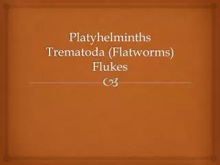
Flat worms, a basic classification Platyhelminthes Trematoda Planaria Aspidobothrea Digenea Monogenea Cestoda
Trematodes • Multicellular eukaryotic helminths • Unsegmented leaf-shaped worms • MONECIOUS except for schistosomes (DIECIOUS)
trematodes or flukes - when they say ‘flat’ worms they mean it • All digenea are parasitic • Small dorso-ventrally flattened worms, unsegmented • No coelom/ body cavity • No blood vessels, simple ladder nervous system
Trematodes or flukes - know your worm • Two suckers (oral and ventral acetabulum): to attach to the host • Oral sucker: mouth • Muscular pharynx permits the worm to pump food into the blind ending gut • Most trematodes are hermaphrodites (self-fertilization occurs)
Trematodes: know your worm • The gut is blind ending, extensive branching • Smooth muscle fibers (longitudinal and cross) run under the tegument and around all the • Body covered with integument with spines - is highly active in nutrient uptake
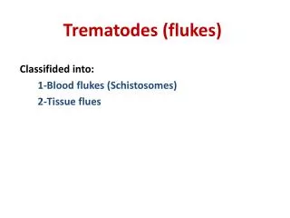
CLASSIFICATION OF TREMATODES • Blood flukes Schistosoma mansoni S. japonicum S. hematobium • Intestinal fluke Fasciolopsis hepatica, F.buski • Liver fluke Clonorchis sinensis, Opistorchis, • Lung fluke Paragonimus westermani.
Life cycle of trematodes Complex life cycle: • Definitive host: Man • Intermediate host: fresh water snail, fish, crab • Some trematodes have 2 or 3 intermediate hosts before the definitive host is reached. • Trematodes inhabit a variety of sites in their hosts: digestive tract, respiratory tract, circulatory system, urinary tract, and reproductive tract.
Life cycles are complex and the fluke passes through numerous stages. • Adult • Egg (or shelled embryo) shed into water • Miracidium: a free swimming, ciliated larva, penetrates a snail (intermediate host) • Sporocyst: reproduces asexually in intermediate host and develops into the next stage called redia. • Rediaproduce more redia or cercariae. Cercariae leave the intermediate host and swim. Then they penetrate the skin of another intermediate host or the definitive host.
Adult Fasciolahepatica fluke (above) Redia (right)
Cercariae : free swimming larvae released by the intermmediate host. Penetrate the skin of the human definitive host • In the definitive host, theyt make their way to their desired home and develop into an adult fluke which reproduces sexually and produces eggs.
Schistosome life cycle.
Pathogenesis: • Local or systemic pathology • Local pathological changes: formation of ulcers/ abcesses- fibrotic changes • Systemic changes: absorption of excretory products
Praziquantel -paralysis of musculature -attachment of phagocytes to parasite and death.
Schistosomiasis • It is believed that Napoleon's army in North Africa was defeated not by the enemy but by infestation of his soldiers with Schistosomal infections.
1 cm The Schistosome
Schistosomiasis (Bilharziasis) • S. hematobium: Africa • S. mansoni : Africa and America • S. japonicum: Far East. • 250 million people are infected
Morphology • Adult worms are 10 to 20 mm long • Male: lamelliform shape with marginal folds
FAVORITE SITES • S. japonicum :VEINS OF GIT • S. mansoni : VEINS OF GIT • S. haematobium : VEINS OF BLADDER
Schistosomiasis • type I and type IV hypersensitivity • collagenase: damage to the vascular endothelium.
Three major disease syndromes occur in schistosomiasis • 1. schistosome dermatitis • 2. acute schistosomias (Katayama fever) • 3. chronic schistosomiasis.
Acute schistosomiasis(Katayama fever) -4 to 8 weeks after primary exposure -cough, hepatosplenomegaly -lymphadenopathy,and eosinophilia
Chronic disease • may appear many years later • japonica and mansoni hepatomegaly splenomegaly portal hypertension esophageal varices
schistosomiasis haematobium inflammation and fibrosis obstruction hydronephrosis uremia
Characteristic eggs: diagnosis Apical spine: S. haematobium Lateral spine: S. mansoni Vestigial spine: S. japonicum
Treatment and control • Praziquantel is effective against all species. • Contaminated water should be avoided. • FILTERING DRINKING WATER • Control measures include sanitary disposal of sewage • No vaccine is available.