Download
1 / 57
590 likes | 739 Views
Neuroscience: The Biological Perspective. Chapter 2. Chapter 2 Menu. How parts of nervous system relate Neurons and nerves and how they work How neurons communicate Neurotransmitters How brain and spinal cord interact Somatic nervous system; interacting with surroundings

E N D
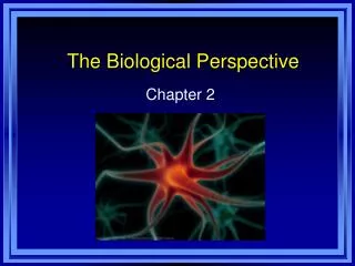
Neuroscience: The Biological Perspective Chapter 2
Chapter 2 Menu • How parts of nervous system relate • Neurons and nerves and how they work • How neurons communicate • Neurotransmitters • How brain and spinal cord interact • Somatic nervous system; interacting with surroundings • Autonomic nervous system and reaction to stress • Study of the brain and how it works • Structures and functions of the bottom part of the brain • Structures that control emotion, learning, memory, motivation • Parts of cortex controlling senses and movement • Parts of cortex responsible for higher forms of thought • Differences between left side and right side of the brain • Hormones interact with nervous system and affect behavior
Parts of nervous system Overview of Nervous System • Nervous System - an extensive network of specialized cells that carry information to and from all parts of the body. • Neuroscience – deals with the structure and function of neurons, nerves, and nervous tissue. • Relationship to behavior and learning. Menu
Neurons and nerves Structure of the Neuron • Neurons - the basic cell that makes up the nervous system and which receives and sends messages within that system. • Parts of a Neuron • Dendrites - branch-like structures that receive messages from other neurons. • Soma - the cell body of the neuron, responsible for maintaining the life of the cell. • Axon - long tube-like structure that carries the neural message to other cells. Menu
Neurons and nerves Menu
Neurons and nerves Other Types of Brain Cells • Glial cells - grey fatty cells that: • provide support for the neurons to grow on and around, • deliver nutrients to neurons, • produce myelin to coat axons, • Myelin - fatty substances produced by certain glial cells that coat the axons of neurons to insulate, protect, and speed up the neural impulse. • clean up waste products and dead neurons. Menu
Neurons and nerves Neurons in the Body • Nerves – bundles of axons in the body that travel together through the body. • Neurilemma – Schwann’s membrane. • Tunnel through which damaged nerve fibers can repair themselves. Menu
LO 2.2 Neurons and nerves Generating the Message: Neural Impulse • Ions – charged particles. • Inside neuron – negatively charged. • Outside neuron – positively charged. • Resting potential - the state of the neuron when not firing a neural impulse. • Action potential - the release of the neural impulse consisting of a reversal of the electrical charge within the axon. • Allows positive sodium ions to enter the cell. • All-or-none - referring to the fact that a neuron either fires completely or does not fire at all. • Return to resting potential. Menu
Sending the Message to Other Cells • Axon terminals - branches at the end of the axon. • Synaptic knob – rounded areas on the end of axon terminals. • Synaptic vesicles - sack-like structures found inside the synaptic knob containing chemicals. • Neurotransmitters - chemical found in the synaptic vesicles which, when released, has an effect on the next cell. • Synapse/synaptic gap - microscopic fluid-filled space between the rounded areas on the end of the axon terminals of one cell and the dendrites or surface of the next cell. • Receptor sites - holes in the surface of the dendrites or certain cells of the muscles and glands, which are shaped to fit only certain neurotransmitters. Menu
Neuron Communication • Neurons must be turned ON and OFF. • Excitatory neurotransmitter - neurotransmitter that causes the receiving cell to fire. • Inhibitory neurotransmitter - neurotransmitter that causes the receiving cell to stop firing. • Chemical substances can affect neuronal communication. • Agonists - mimic or enhance the effects of a neurotransmitter on the receptor sites of the next cell, increasing or decreasing the activity of that cell. • Antagonists - block or reduce a cell’s response to the action of other chemicals or neurotransmitters. Menu
Neurotransmitters • Types of neurotransmitters Menu
Neurotransmitters Cleaning up the Synapse • Reuptake - process by which neurotransmitters are taken back into the synaptic vesicles. • Enzyme - a complex protein that is manufactured by cells. • One type specifically breaks up acetylcholine because muscle activity needs to happen rapidly, so reuptake would be too slow. Menu
Brain and spinal cord Central Nervous System • Central nervous system (CNS) - part of the nervous system consisting of the brain and spinal cord. • Spinal cord - a long bundle of neurons that carries messages to and from the body to the brain that is responsible for very fast, lifesaving reflexes. Menu
The Reflex Arc: Three Types of Neurons • Sensory neuron - a neuron that carries information from the senses to the central nervous system. • Also called afferent neuron. • Motor neuron - a neuron that carries messages from the central nervous system to the muscles of the body. • Also called efferent neuron. • Interneuron - a neuron found in the center of the spinal cord that receives information from the sensory neurons and sends commands to the muscles through the motor neurons. • Interneurons also make up the bulk of the neurons in the brain. Menu
Somatic nervous system ----- Autonomic nervous system Peripheral Nervous System • Peripheral nervous system (PNS) - all nerves and neurons that are not contained in the brain and spinal cord but that run through the body itself; divided into the: • Somatic nervous system • Autonomic nervous system Menu
Somatic Nervous System • Soma = body. • Somatic nervous system - division of the PNS consisting of nerves that carry information from the senses to the CNS and from the CNS to the voluntary muscles of the body. • Sensory pathway - nerves coming from the sensory organs to the CNS consisting of sensory neurons. • Motor pathway - nerves coming from the CNS to the voluntary muscles, consisting of motor neurons. Menu
Autonomic Nervous System • Autonomic nervous system (ANS) - division of the PNS consisting of nerves that control all of the involuntary muscles, organs, and glands sensory pathway nerves coming from the sensory organs to the CNS consisting of sensory neurons. • Sympathetic division (fight-or-flight system) - part of the ANS that is responsible for reacting to stressful events and bodily arousal. • Parasympathetic division - part of the ANS that restores the body to normal functioning after arousal and is responsible for the day-to-day functioning of the organs and glands. Menu
Peeking Inside the Brain • Clinical studies • Deep lesioning - insertion of a thin, insulated wire into the brain through which an electrical current is sent that destroys the brain cells at the tip of the wire. • Electrical stimulation of the brain (ESB) – milder electrical current that causes neurons to react as if they had received a message. • Human brain damage. • Electroencephalograph (EEG) - machine designed to record the brain wave patterns produced by electrical activity of the surface of the brain. Menu
Peeking Inside the Brain • Magnetic resonance imaging (MRI) - brain-imaging method using radio waves and magnetic fields of the body to produce detailed images of the brain. • Functional MRI (fMRI) – computer makes a sort of “movie” of changes in the activity of the brain using images from different time periods. • Computed tomography (CT) - brain-imaging method using computer controlled X-rays of the brain. • Positron emission tomography (PET) - brain-imaging method in which a radioactive sugar is injected into the subject and a computer compiles a color-coded image of the activity of the brain with lighter colors indicating more activity. Menu
Structures of the bottom part of brain The Brain Stem • Medulla - the first large swelling at the top of the spinal cord, forming the lowest part of the brain, which is responsible for life-sustaining functions such as breathing, swallowing, and heart rate. • Pons - the larger swelling above the medulla that connects the top of the brain to the bottom and that plays a part in sleep, dreaming, left–right body coordination, and arousal. Menu
Structures of the bottom part of brain The Brain Stem • Reticular formation (RF) - an area of neurons running through the middle of the medulla and the pons and slightly beyond that is responsible for selective attention. • Cerebellum - part of the lower brain located behind the pons that controls and coordinates involuntary, rapid, fine motor movement. Menu
Structures controlling emotion, learning, memory, and motivation Structures Under the Cortex • Limbic system - a group of several brain structures located under the cortex and involved in learning, emotion, memory, and motivation. • Thalamus - part of the limbic system located in the center of the brain, this structure relays sensory information from the lower part of the brain to the proper areas of the cortex and processes some sensory information before sending it to its proper area. • Olfactory bulbs - two projections just under the front of the brain that receive information from the receptors in the nose located just below. Menu
Structures controlling emotion, learning, memory, and motivation Structures Under the Cortex • Limbic system (continued) • Hypothalamus - small structure in the brain located below the thalamus and directly above the pituitary gland, responsible for motivational behavior such as sleep, hunger, thirst, and sex. • Sits above and controls the pituitary gland (master endocrine gland). • Hippocampus - curved structure located within each temporal lobe, responsible for the formation of long-term memories and the storage of memory for location of objects. • Amygdala - brain structure located near the hippocampus, responsible for fear responses and memory of fear. Menu
Structures controlling emotion, learning, memory, and motivation Cortex • Cortex - outermost covering of the brain consisting of densely packed neurons, responsible for higher thought processes and interpretation of sensory input. • Corticalization – wrinkling of the cortex. • Allows a much larger area of cortical cells to exist in the small space inside the skull. Menu
Structures controlling emotion, learning, memory, and motivation Human cortex compared to various animal species Menu
Parts of cortex controlling senses and movement Cerebral Hemispheres • Cerebral hemispheres - the two sections of the cortex on the left and right sides of the brain. • Corpus callosum - thick band of neurons that connects the right and left cerebral hemispheres. Menu
Parts of cortex controlling senses and movement Four Lobes of the Brain • Occipital lobe - section of the brain located at the rear and bottom of each cerebral hemisphere containing the visual centers of the brain. • Primary visual cortex – processes visual information from the eyes. • Visual association cortex – identifies and makes sense of visual information. • Parietal lobes - sections of the brain located at the top and back of each cerebral hemisphere containing the centers for touch, taste, and temperature sensations. • Somatosensory cortex - area of neurons running down the front of the parietal lobes responsible for processing information from the skin and internal body receptors for touch, temperature, body position, and possibly taste. Menu
Parts of cortex controlling senses and movement Four Lobes of the Brain • Temporal lobes - areas of the cortex located just behind the temples containing the neurons responsible for the sense of hearing and meaningful speech. • Primary auditory cortex – processes auditory information from the ears. • Auditory association cortex – identifies and makes sense of auditory information. • Frontal lobes - areas of the cortex located in the front and top of the brain, responsible for higher mental processes and decision making as well as the production of fluent speech. • Motor cortex - section of the frontal lobe located at the back, responsible for sending motor commands to the muscles of the somatic nervous system. Menu
The homunculus is commonly used today in scientific disciplines, such as psychology, as a teaching or memory tool to describe the distorted scale model of a human drawn or sculpted to reflect the relative space human body parts occupy on the somatosensory cortex (sensory homunculus) and the motor cortex (motor homunculus). Menu
Parts of cortex responsible for higher thought Association Areas of Cortex • Association areas - areas within each lobe of the cortex responsible for the coordination and interpretation of information, as well as higher mental processing. • Broca’s aphasia - condition resulting from damage to Broca’s area (usually in left frontal lobe), causing the affected person to be unable to speak fluently, to mispronounce words, and to speak haltingly. • Wernicke’s aphasia - condition resulting from damage to Wernicke’s area (usually in left temporal lobe), causing the affected person to be unable to understand or produce meaningful language. • Spatial neglect - condition produced by damage to the association areas of the right hemisphere resulting in an inability to recognize objects or body parts in the left visual field. Menu
Parts of cortex responsible for higher thought Spatial neglect Menu
Left side and right side of brain Split Brain Research • Cerebrum - the upper part of the brain consisting of the two hemispheres and the structures that connect them. • Split brain research • Study of patients with severed corpus callosum. • Involves sending messages to only one side of the brain. • Demonstrates right and left brain specialization. Menu
Left side and right side of brain Split-brain subjects stared at a dot and viewed a composite of two faces (A). When asked what they saw, subjects chose the child—the image sent to the verbal left hemisphere (B). But when subjects pointed to the face with the left hand, they chose the woman with glasses—whose image was received by the right hemisphere (C) (Levy et al., 1983). Menu
Left side and right side of brain Language is primarily a left hemisphere activity for most individuals Menu
Left side and right side of brain Results of Split Brain Research • Left side of the brain: • seems to control language, writing, logical thought, analysis, and mathematical abilities, • processes information sequentially, • can speak. • Right side of the brain • controls emotional expression, spatial perception, recognition of faces, patterns, melodies, and emotions, • processes information globally, • cannot speak. Menu