Download
1 / 34
340 likes | 520 Views
Recombinant DNA Technology (8). Cloning Eukaryotic DNAs in Phage Genomes Recombinant DNA molecules are packaged into lambda phage heads. Lambda phage can take inserts up to a size of 25 kb. Sites of phage infection are identified as plaques in a bacterial “lawn”.

E N D
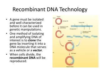
Recombinant DNA Technology (8) • Cloning Eukaryotic DNAs in Phage Genomes • Recombinant DNA molecules are packaged into lambda phage heads. • Lambda phage can take inserts up to a size of 25 kb. • Sites of phage infection are identified as plaques in a bacterial “lawn”. • DNA fragments are identified by the same techniques as those used for cloning of recombinant plasmids.
Protocol for cloning eukaryotic DNA fragments in lambda phage
18.14 Enzymatic Amplification of DNA by PCR (1) • Polymerase chain reaction (PCR) is a technique to amplify specific DNA fragments. • It uses a very small amount of template. • Utilizes a heat-stable DNA polymerase (Taq polymerase) from bacteria living in hot springs. • Uses repeated cycles of denaturation, DNA replication, and cooling to double the amount of DNA during each cycle. • Uses an automated thermal cycler.
Enzymatic Amplification of DNA by PCR (2) • Applications of PCR • Amplifying DNA for cloning or analysis—such as in criminal investigations and fossil analysis. • Testing for the presence of specific DNA sequences • Comparing DNA molecules • Quantifying DNA or RNA templates
18.15 DNA Sequencing (1) • Techniques developed in the 1970s are widely used for sequencing nucleic acids. • The Sanger-Coulson dideoxy method became the most widely used: • Four samples of identical single-stranded DNA molecules are obtained.
DNA Sequencing (2) • Dideoxy sequencing (continued) • DNA of each sample is incubated with a primer, DNA polymerase, four dNTPs, and a low concentration of ddNTPs (dideoxyribonucleoside triphosphates), different one in each sample. • DNA fragments of different lengths are synthesized in each sample, with synthesis terminating where ddNTP has been randomly incorporated.
DNA Sequencing (3) • Dideoxy sequencing (continued) • DNA fragments are separated by gel electrophoresis and the DNA sequence is read form the gel bands. • The amino acid sequence of the protein is deduced from the nucleotide sequence.
DNA Sequencing (4) • Next-generation sequencing (NGS) is based on polymerase-dependent DNA synthesis but does not depend upon premature termination. • It identifies the individual nucleotides as they are being incorporated by the polymerase in real time.
18.16 DNA Libraries (1) • DNA libraries are often produced from DNA cloning: • Genomic libraries are produced from DNA extracted from nuclei and contain all DNA sequences of the species. • cDNA libraries are derived from DNA copies of an RNA population.
DNA Libraries (2) • Genomic Libraries • DNA fragments for the library can be obtained by cutting genomic DNA with restriction enzymes. • Cleaving of genomic DNA is random, which generates overlapping fragments. • Overlapping fragments are useful for chromosome walking, to study linked sequences in an extended region of a chromosome.
DNA Libraries (3) • Cloning Larger DNA Fragments in Specialized Cloning Vectors • A yeast artificial chromosome (YAC) can accommodate large (up to 1000 kb) DNA inserts. • A bacterial artificial chromosome (BAC) accepts DNA inserts of up to 500 kb, and can be quickly grown to large numbers.
DNA Libraries (4) • cDNA Libraries • The isolation of genomic fragments allows study of the genome. • Coding regions of a gee can be studied using cDNAs, synthesized by reverse transcriptase using mRNA as a template. • Allow foreign DNA to be transcribed and translated during the infection process.
18.17 DNA Transfer into Eukaryotic Cells and Mammalian Embryos (1) • DNA incorporation into the genome of a nonreplicating virus is called transduction. • DNA introduced into cultured cells is called transfection. • The gene whose role is being investigated after transfection is called a transgene.
DNA Transfer into Eukaryotic Cells and Mammalian Embryos (2) • A direct way to introduce foreign genes into a cell is by microinjection of DNA directly into the cell nucleus. • Animals that have been genetically engineered to that their chromosomes have foreign genes are called transgenic animals.
DNA Transfer into Eukaryotic Cells and Mammalian Embryos (3) • Transgenic Plants and Animals • Transgenic organisms allow scientists to determine the effects of overexpression of a particular DNA sequence. • Genetic engineering can produce animal models used to study human diseases. • The main goal of genetic engineering in plants is to improve the efficiency of both photosynthesis and nitrogen fixation.
18.18 Determining Eukaryotic Gene Function by Gene Elimination (1) • In Vitro Mutagenesis • Site-directed mutagenesis (SDM) allows making small changes in a DNA sequence. • SDM is accomplished by synthesizing a DNA containing the desired change and allowing it to hybridize to a single-stranded normal DNA. • The polymerase elongates the replicates DNA adding nucleotides complementary to the normal DNA.
Determining Eukaryotic Gene Function by Gene Elimination (2) • Knockout Mice • Knockout mice are obtained by transfecting embryonic stem cells, introducing them into an embryo, and implanting the embryo into a female mouse. • Germ cells containing the knockout mutation are heterozygous, which can be used to obtain a homozygous mutant phenotype. • Genes can be assessed for their functions.
Determining Eukaryotic Gene Function by Gene Elimination (3) • RNA Interference • Specific mRNAs can be degraded in vivo by treating with small double-stranded siRNA containing part of the sequence of the target mRNA. • Cells treated with RNAi cannot make the protein encoded in the target mRNA. • Libraries containing thousands of siRNAs are available for the study of gene function.
18.19 The Use of Antibodies (1) • Antibodies are highly specific proteins produced by lymphoid tissues in response to the presence of foreign materials. • Preparation of antibodies: • A population of polyclonal antibodies can be obtained by repeated injections of a purified antigen into an animal. • The blood of the animal serves as a source of an antiserum.
The Use of Antibodies (2) • Preparation of antibodies (continued): • A monoclonal antibody is produced by descendants of a single antibody-producing cell: • Antibody-producing cells do not grow and divide in culture. • Fusion of a normal antibody-producing lymphocyte and a malignant myeloma cell will create a viable hybridoma cell that can produce large amounts of a monoclonal antibody.
The Use of Antibodies (3) • Antibodies can be conjugated with a fluorescent substance that allows visualization of antigens. • In direct immunofluorescence, antibodies with bound fluorescent molecules bind to antigens and can be visualized with a fluorecence microscope. • In indirect immunofluorescence, cells are incubated with unlabeled antibodies, and then with labeled 2˚ antibody against the 1˚ antibody.