Download
1 / 19
200 likes | 396 Views
Diagnosis of TB. Learning Objectives. List the 4 principle components of a TB evaluation Describe the criteria which differentiate PTB+ from PTB- Describe the 3 major indications for culture and DST. Common Sites of TB Disease. Lungs Pleura Central nervous system Lymphatic system

E N D
Learning Objectives • List the 4 principle components of a TB evaluation • Describe the criteria which differentiate PTB+ from PTB- • Describe the 3 major indications for culture and DST
Common Sites of TB Disease • Lungs • Pleura • Central nervous system • Lymphatic system • Genitourinary systems • Bones and joints • Disseminated (miliary TB)
Systemic Symptoms of TB • Fever • Chills • Night sweats • Appetite loss • Weight loss • Fatigue
Evaluation for TB • HIV test • Medical history • Physical examination • Bacteriologic or histologic exam • (Chest radiograph if indicated)
Medical History • HIV status • Symptoms of disease • History of TB exposure, infection, or disease • Past TB treatment • Demographic risk factors for TB • Other medical conditions that increase risk for TB disease (e.g., diabetes)
Symptoms of Pulmonary TB • Productive, prolonged cough • (duration of 2-3 weeks) • Chest pain • Hemoptysis (bloody sputum) • Signs may vary based on HIV status
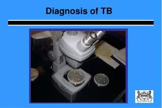
Specimen Collection Procedure • Obtain 3 sputum specimens for smear examination • and culture • Spot, first morning, spot • Follow infection control precautions during • specimen collection
Sputum Smear Examination • Specimens should be sent to the lab immediately to preserve the quality of the specimens • Always aim for three specimens at each exam • Always store at a cool temperature and away from sunlight to preserve the quality of specimens • 3 respiratory specimens will detect 90% of smear-positive cases
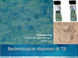
AFB smear-microscopy Acid-fast bacilli (AFB) (shown in red) are tubercle bacilli
Smear-positive PTB vs.Smear-negative PTB- • PTB+ (Pulmonary TB smear-positive) • One AFB-positive smear; i.e. any patient with at least one positive smear result (irrespective of quantity of AFBs seen on microscopy) • Recommendations to improve the diagnosis of smear negative pulmonary and extrapulmonary TB among adults in HIV prevalent and resource constrained settings. • Draft for discussion by Strategic and Technical Advisory Group of Stop TB Department of WHOJune 2006
Smear-positive PTB vs.Smear-negative PTB- • PTB- (smear-negative) Any pulmonary TB case that does not meet the definition of being smear-positive. This includes: 1. Patients with three negative smear results and radiological findings and doctor’s decision to treat for TB 2. Patients with negative smear results and a positive culture result for M. tuberculosis 3. Patients who are unable to produce sputum and with highly suspicious radiological and clinical findings and doctor's decision to treat for TB
Other Acid Fast Bacilli • Mycobacteria other than those comprising the M. tuberculosis complex are called Non-Tuberculous Mycobacteria (“NTM”) or Mycobacteria Other Than Tuberculosis (“MOTT”). • These mycobacteria may cause pulmonary disease resembling TB. Increasingly, cases from these organisms are being reported in patients with weakened immune systems, especially due to HIV. • It is important to note that infection with MOTT also may produce AFB-positive sputum smear results and positive Mantoux skin test readings mimicking M. tuberculosis. Culture can distinguish between M. tuberculosis and MOTT. Disease due to MOTT is usually unresponsive to first-line anti-TB drugs.
Chest Radiograph • Diagnosis of PTB solely on basis of CXR not encouraged • May have unusual appearance in • HIV-positive persons • CXR is helpful in HIV+, smear- negative patients • Cannot confirm diagnosis of TB Arrow points to cavity in patient's right upper lobe.
Cultures • Should be requested for ALL retreatment patients • Relapse • Failure • Return after default • Culture is indicated for • New and retreatment PTB cases still smear- positive at end of intensive phase • Symptomatic contacts of known MDR cases Colonies of M. tuberculosis growing on media
Diagnosis in Children • Patient history • Contact to PTB+ • Symptoms consistent with TB • HIV test • Clinical Exam • TST • Bacteriological confirmation • Investigations for PTB and EPTB Guidance of National Tb Programmes for the Management of TB in Children WHO/HTM/TB/2006.371
Key Risk Factors in Children Risk Factors For Children Include: • Household contact with a newly diagnosed smear-positive case • Age less than 5 years • HIV infection • Severe malnutrition.
Key Features of TB in Children The presence of three or more of the following should strongly suggest a diagnosis of TB: • Chronic symptoms suggestive of TB • Physical signs highly of suggestive of TB • A positive tuberculin skin test • Chest X-ray suggestive of TB (The presentation in infants may be more acute, resembling acute severe pneumonia and should be suspected when there is a poor response to antibiotics. In such situations, there is often an identifiable source case, usually the mother.)