Download
1 / 22
220 likes | 261 Views
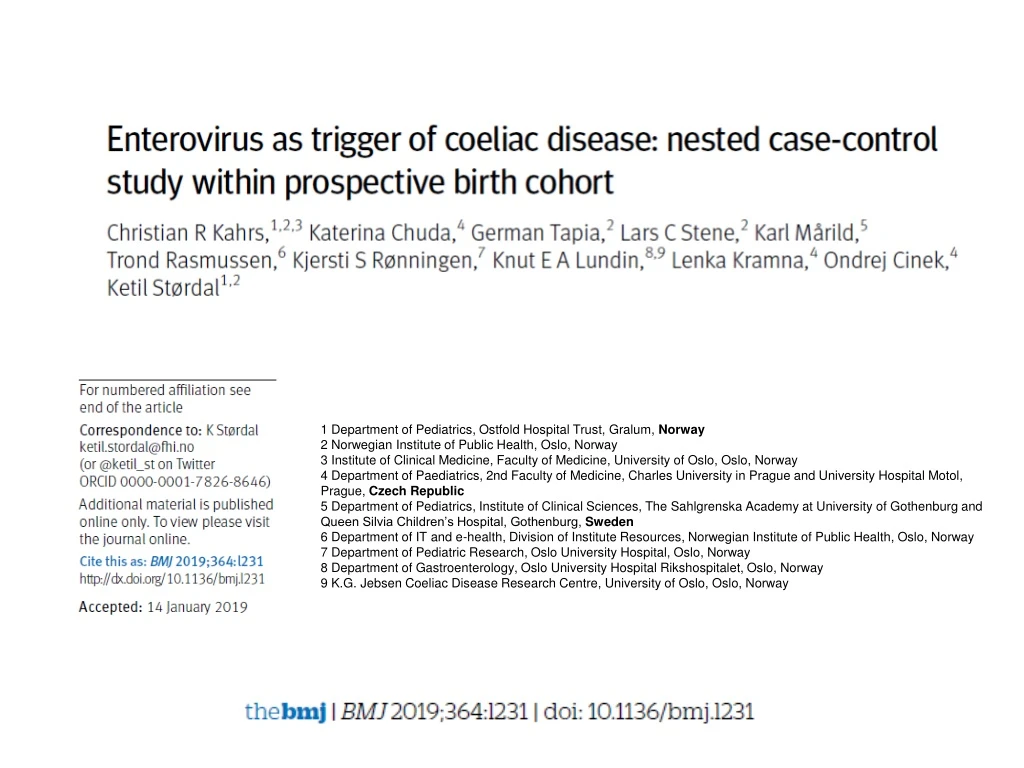
This study investigates the impact of gastrointestinal viral infections, specifically enterovirus and adenovirus, on the development of coeliac disease. By analyzing a longitudinal birth cohort, the research aims to determine if these common viruses play a role in triggering coeliac disease. Understanding potential triggers is crucial for early detection and prevention strategies.

E N D
1 Department of Pediatrics, Ostfold Hospital Trust, Gralum, Norway 2 Norwegian Institute of Public Health, Oslo, Norway 3 Institute of Clinical Medicine, Faculty of Medicine, University of Oslo, Oslo, Norway 4 Department of Paediatrics, 2nd Faculty of Medicine, Charles University in Prague and University Hospital Motol, Prague, Czech Republic 5 Department of Pediatrics, Institute of Clinical Sciences, The Sahlgrenska Academy at University of Gothenburg and Queen Silvia Children’s Hospital, Gothenburg, Sweden 6 Department of IT and e-health, Division of Institute Resources, Norwegian Institute of Public Health, Oslo, Norway 7 Department of Pediatric Research, Oslo University Hospital, Oslo, Norway 8 Department of Gastroenterology, Oslo University Hospital Rikshospitalet, Oslo, Norway 9 K.G. Jebsen Coeliac Disease Research Centre, University of Oslo, Oslo, Norway
BACKGROUND (I) • Coeliac disease is an immune mediated disease believed to result from gluten intake and unknown environmental trigger factors in genetically susceptible individuals.1 • Coeliac disease develops almost exclusively in people with the HLA-DQ2 (DQA1*05:01- QB1*02:01) or HLA-DQ8 (DQA1*03-DQB1*03:02) haplotype. The HLA-DQ2 and HLA-DQ8 haplotypes also occur in about 40% of the general population. • Although non-HLA genetic variants have also been identified, the predictive value of these variants, even in combination, is limited.2 3 1 Green PH, Cellier C. Celiac disease. N Engl J Med 2007;357:1731-43.doi:10.1056/NEJMra071600 2 Romanos J, Rosen A, Kumar V, et al, PreventCD Group. Improving coeliac disease risk prediction by testing non-HLA variants additional to HLA variants. Gut 2014;63:415-22. doi:10.1136/ gutjnl-2012-304110 3 Marild K, Vistnes M, Tapia G, et al. Midpregnancy and cord blood immunologic biomarkers, HLA genotype, and pediatric celiac disease. J Allergy Clin Immunol 2017;139:1696-8. doi:10.1016/j. jaci.2016.10.016
BACKGROUND (II) • Both experimental studies and epidemiological studies based on parental reporting of illness suggest a role for infections in the development of coeliac disease, particularly gastrointestinal infections.6-9 • Gastrointestinal infections are common in childhood and may impair the mucosal barrier for transfer of dietary proteins as gluten regardless of the presence of clinical symptoms.10 11 • The only prospective study on coeliac disease and viral infection suggested that frequent rotavirus infections might increase the risk of development of coeliac disease antibodies in a cohort at high risk.12 • Other studies with retrospective designs have studied adenovirus, enterovirus, and orthoreovirus as potential triggers of coeliac disease with conflicting or inconclusive results.9 13-15 6 Abadie V, Sollid LM, Barreiro LB, Jabri B. Integration of genetic and immunological insights into a model of celiac disease pathogenesis. Annu Rev Immunol 2011;29:493-525. doi:10.1146/annurevimmunol- 040210-092915 7 Marild K, Kahrs CR, Tapia G, Stene LC, Stordal K. Infections and risk of celiac disease in childhood: a prospective nationwide cohort study. Am J Gastroenterol 2015;110:1475-84. doi:10.1038/ajg.2015.287 8 Kemppainen KM, Lynch KF, Liu E, et al, TEDDY Study Group. Factors That Increase Risk of Celiac Disease Autoimmunity After a Gastrointestinal Infection in Early Life. Clin Gastroenterol Hepatol 2017;15:694-702.e5. doi:10.1016/j.cgh.2016.10.033 9 Bouziat R, Hinterleitner R, Brown JJ, et al. Reovirus infection triggers inflammatory responses to dietary antigens and development of celiac disease. Science 2017;356:44-50. doi:10.1126/science. aah5298 10 Webb A, Starr M. Acute gastroenteritis in children. Aust Fam Physician 2005;34:227-31. 11 Schumann M, Siegmund B, Schulzke JD, Fromm M. Celiac Disease: Role of the Epithelial Barrier. Cell Mol Gastroenterol Hepatol 2017;3:150-62. doi:10.1016/j.jcmgh.2016.12.006 12 Stene LC, Honeyman MC, Hoffenberg EJ, et al. Rotavirus infection frequency and risk of celiac disease autoimmunity in early childhood: a longitudinal study. Am J Gastroenterol 2006;101:2333-40. doi:10.1111/j.1572-0241.2006.00741.x 13 Kagnoff MF, Paterson YJ, Kumar PJ, et al. Evidence for the role of a human intestinal adenovirus in the pathogenesis of coeliac disease. Gut 1987;28:995-1001. doi:10.1136/gut.28.8.995 14 Mahon J, Blair GE, Wood GM, Scott BB, Losowsky MS, Howdle PD. Is persistent adenovirus 12 infection involved in coeliac disease? A search for viral DNA using the polymerase chain reaction. Gut 1991;32:1114-6. doi:10.1136/gut.32.10.1114 15 Mercalli A, Lampasona V, Klingel K, et al. No evidence of enteroviruses in the intestine of patients with type 1 diabetes. Diabetologia 2012;55:2479-88. doi:10.1007/s00125-012-2591-4
AIMS AND METHODS • In this study, we approached the question of potential gastrointestinal triggers by using a longitudinal birth Cohort analysis of the most frequently occurring viruses: enterovirus and adenovirus. • We aimed to test whether the presence of human enterovirus and adenovirus in monthly faecal samples was more common before development of coeliac disease antibodies in cases subsequently diagnosed as having coeliac disease compared with children not developing the disease. • Nested case-control study of coeliac disease within a birth cohort of children with the HLA-DQ2/DQ8 genotype
STUDY DESIGN Fig 1 | Enrolment of study sample. “Invited” children are those who were still actively participating (delivering samples, questionnaires) at start of coeliac disease sub-study and were invited to participate; “participated” are those who consented to participate and were screened for coeliac disease antibodies. *Two cases were excluded from analyses, one owing to missing stool samples and one owing to incorrect use of diagnostic criteria. †Children with positive coeliac disease antibodies at first screening test but for whom follow-up did not confirm coeliac disease diagnosis (n=10) were excluded from further analyses; control children with high antibody titres (>10 times cut-off) (n=2) in old samples but with normal values at screening were excluded and replaced by new controls selected using same matching criteria. ‡One matched control was excluded from analysis owing to missing stool samples
TIMELINE OF THE STUDY We collected plasma samples at age 3, 6, 9, and 12 months and annually thereafter. Monthly stool samples collected at age 3-36 months were diluted in preservation buffer, centrifuged, and supernatant separated.
CASE DEFINITION AND SELECTION OF MATCHED CONTROLS • We diagnosed coeliac disease according to the European Society for Paediatric Gastroenterology Hepatology and Nutrition (ESPGHAN) 2012 criteria • 25 cases (16 girls and 9 boys; 11% of participants) with coeliac disease matched to two controls each. Matching was done for duration of follow-up, date of birth, and county of residence. • Mean age at end of follow-up was 9.9 (SD 1.6) years
ESPGHAN Guidelines for Diagnosis of Coeliac Disease JPGN Volume 54, Number 1, January 2012
ESPGHAN Guidelines for Diagnosis of Coeliac Disease JPGN Volume 54, Number 1, January 2012
CASE DEFINITION AND SELECTION OF MATCHED CONTROLS • We then determined the time interval when cases seroconverted for coeliac disease markers by retrospectively analysing biobanked plasma samples that had been collected longitudinally since age 3 months, searching for the last sample that was negative for the serological markers and the first sample indicative of coeliac disease. • Of the 25 case control groups, 15 had this seroconversion period covered by monthly stool sampling, whereas the remaining 10 seroconverted after the collection of stools was terminated (after 36 months of age).
DETECTION OF ENTEROVIRUS AND ADENOVIRUS IN STOOL SAMPLES • All available faecal samples from cases and controls were subjected to RNA and DNA extraction using Qiagen chemistry (Qiagen, Hilden, Germany). • We tested enterovirus by quantitative real time polymerase chain reaction (PCR).23 • Adenovirus was tested using a previously published real time PCR assay24 • The threshold of enterovirus and adenovirus positivity was set to 10 copies/μL nucleic acid. 23 Honkanen H, Oikarinen S, Pakkanen O, et al. Human enterovirus 71 strains in the background population and in hospital patients in Finland. J Clin Virol 2013;56:348-53. doi:10.1016/j. jcv.2012.11.018 24 Claas EC, Schilham MW, de Brouwer CS, et al. Internally controlled real-time PCR monitoring of adenovirus DNA load in serum or plasma of transplant recipients. J Clin Microbiol 2005;43:1738-44. doi:10.1128/JCM.43.4.1738-1744.2005
DETECTION OF ENTEROVIRUS AND ADENOVIRUS IN STOOL SAMPLES • We tested 2161 stool samples, of which 2135 and 2006 provided data on quantity of enterovirus and adenovirus, respectively. All samples were tested blinded as to the case-control status. • All samples with more than 100 copies/μL of enterovirus were subjected to genotyping with Sanger next generation amplicon sequencing of PCR amplicons of the VP1 gene segment informative for the virus type. • Samples with more than 10 copies/μL of adenovirus were genotyped by a similar protocol amplifying the seventh hypervariable region of its hexon gene.
STATISTICAL ANALYSIS • We analysed the association of virus with coeliac disease primarily by using a mixed effects logistic regression model • We used faecal sample virus positivity as the dependent variable (separate models for enterovirus and adenovirus) and case-control status as an independent variable. • The odds ratio for coeliac disease status is then interpreted as the odds of a faecal sample being positive for virus given that it came from a child who later developed coeliac disease, compared with the same odds for virus positivity for samples from matched controls. • Potential predictors of viral infection and coeliac disease as covariates in the main regression model: sex, age, age squared, season of sample collection, number of siblings (categorised as none, 1, or ≥2), and family history of coeliac disease.
STATISTICAL ANALYSIS • Exposure defined as number of infectious episodes, counting a sequence of consecutively virus positive faecal samples as a single episode (a negative stool sample is required before a new episode is defined). • We speculated that a higher quantity of enterovirus, longer duration of viral shedding, or symptomatic infections would have a greater effect on development of coeliac disease. • Additionally, we adjusted the primary analysis for the timing of introduction of gluten and breast feeding.
RESULTS • We detected enterovirus in 370 (17%) of 2135 stool Samples, with 73 children having at least one positive sample. • The frequency of enterovirus positive stool samples before development of coeliac disease antibodies was 84/429 (20%) in cases and 129/855 (15%) in matched controls (adjusted odds ratio 1.49, 95% confidence interval 1.07 to 2.06; P=0.02) • The adjusted odds ratios were: • 2.11 (1.24 to 3.60; P=0.01) for high quantity samples (>100 000 copies/μL), • 2.16 (1.16 to 4.04; P=0.02) for long lasting infections (more than two months), • 1.27 (0.87 to 1.86; P=0.21) for infectious episodes (consecutive positive samples counted as a single episode) • The frequency of enterovirus in stool samples during or after development of coeliac disease antibodies was not associated with coeliac disease
ENTEROVIRUS AND CELIAC DISEASE • Enterovirus infections after the first year of life showed increased estimates, whereas infections from age 3 to 6 months or from 6 to 12 months did not. • Enterovirus infections after introduction of gluten were associated with coeliac disease, whereas infections before or at the time of gluten introduction were not • Similarly, infections after the end of breast feeding were associated with coeliac disease, but enterovirus infections during breast feeding were not. • We found no association between reported infectious symptoms and coeliac disease or between infectious symptoms and enterovirus positivity
STRENGTHS OF THE STUDY • First population based study on viruses in stool samples collected longitudinally before development of coeliac disease antibody markers in children later diagnosed as having coeliac disease • We used a PCR technique, which is a highly sensitive method for detection of virus locally in the gut but does not measure the immune response as serological testing does • We used a quantitative assay that enabled us to follow the dynamics of the viral load. • Regular monthly stool sampling and high completeness were important strengths, because duration of viral shedding is expected to be around three to four weeks
LIMITATIONS OF THE STUDY • Participants were followed up for about 10 years, and some children (particularly among those with the shortest followup) would be likely to be diagnosed as having coeliac disease after that age • Limited sample size for some of the sub-analyses could have led to spurious associations • Loss to follow-up and modest response rate is inevitable in most cohort studies and also raises the possibility of selection bias
CONCLUSIONS • Although the effect sizes are relatively small, this study suggests that infections with enterovirus in early life could be one among several key risk factors for development of a disease with lifelong consequences • Rather than a specific enterovirus species or type driving the association, several enterovirus types, high titre, and long duration infections in the period after introduction of gluten were involved • If enterovirus is confirmed as a trigger factor, vaccination could reduce the risk of development of coeliac disease • Given the limited number of cases, we call for corroboration in similar studies and preferably interventional studies to reach conclusions about causality.