Download
1 / 44
440 likes | 605 Views
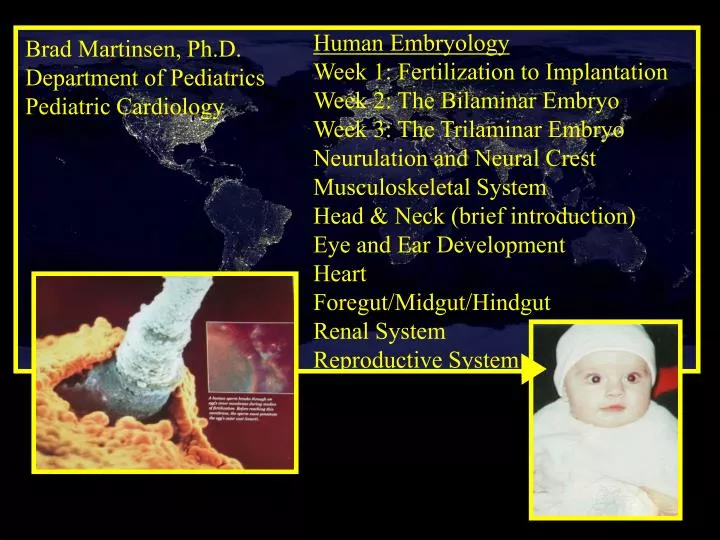
Human Embryology Week 1: Fertilization to Implantation Week 2: The Bilaminar Embryo Week 3: The Trilaminar Embryo Neurulation and Neural Crest Musculoskeletal System Head & Neck (brief introduction) Eye and Ear Development Heart Foregut/Midgut/Hindgut Renal System Reproductive System.

E N D
Human Embryology Week 1: Fertilization to Implantation Week 2: The Bilaminar Embryo Week 3: The Trilaminar Embryo Neurulation and Neural Crest Musculoskeletal System Head & Neck (brief introduction) Eye and Ear Development Heart Foregut/Midgut/Hindgut Renal System Reproductive System Brad Martinsen, Ph.D. Department of Pediatrics Pediatric Cardiology
1.Reading assignments for first exam:Human Embryology, Larsen, 3rd & 2nded. Larson pp1-67 (1-63) Larson pp 80-93, 98-102 (73-87, 92-96) Larson pp 315-335 (311-329)2. Exam #1--> Handouts are a complete review of the reading and what I will say in lecture. Use reading assignments to clarify anything said in lecture or in the handout. Not all detail in the reading assignments will be on the exam.
First Days of Development Day 0-5 Compaction starts
Review Question. Which of the following is the origin of the mitochondrial DNA of all human Adult Cells? A) Paternal only B) Maternal only C) A combination of paternal and maternal D) Either paternal or maternal E) Unknown origin
Review Question. Which of the following is the origin of the mitochondrial DNA of all human Adult Cells? A) Paternal only B) Maternal only C) A combination of paternal and maternal D) Either paternal or maternal E) Unknown origin
Days 12-13 Chorion membrane consists of?
Day 14-15 Primary ectoderm: primordial germ cells, endothelial cells, and hematopoietic stem cells. Extraembryonic mesoderm of the yolk sac wall is a major site of hematopoiesis. Chorion membrane consists of?
Origin of the germ lineA. Migration of the primordial germ cells.B. Migration into posterior body wall. 2 Weeks ->ovarian follicle cells 10th Thoracic Vertebrae 4-6 weeks ->Sertoli cells Teratoma:
Clinical ApplicationHuman chorionic gonadotropin (hCG) • hCG is a 57,000 MW glycoprotein with two subunits • (alpha and beta) produced by the syncytiotrophoblast. • Enters the maternal blood circulation. • Prevents degeneration of the corpus luteum. • Stimulates production of progesterone in the corpus luteum • and chorion, which sustains the placenta. • Can be assayed in maternal blood at day 8 after fertilization • And in maternal urine at day 10. This is the basis of early diagnosis of pregnancy.
Clinical ApplicationComplete hydatidiform mole Persistent trophoblastic disease Metastatic choriocarcinoma
IV. Clinical Applications.F. Genomic Imprinting. 1. Cytogenetic analysis of hydatidiform moles suggests Paternal genetic complement->placental development. Maternal genetic complement->embryo development. 2. Methylation of DNA is a mechanism that leads to independent expression of maternal and paternal genomes during early development. Female germ line highly methylated. 3. Example of pattern of inheritance: Father->Prader-Willi Syndrome Mother->Angelman Syndrome 4. Severity and age of onset of several genetic diseases also differ on inheritance pattern.
Week Three.I. Gastrulation and somite/Nt development.A. Primitive Streak.
I. Gastrulation and somite/Nt development.B. Process of Gastrulation.
I. Gastrulation and somite/Nt development.B. Process of Gastrulation.
I. Gastrulation and somite/Nt development.B. Process of Gastrulation.
I. Gastrulation and somite/Nt development.B. Process of Gastrulation.
I. Gastrulation and somite/Nt development.C. Paraxial, intermediate, and lateral plate mesoderm. Axial skel. Vol muscl. Dermis Splanchnopleuric Somatopleuric mesoderm Urinary system Genital system
I. Gastrulation and somite/Nt development.C. Paraxial, intermediate, and lateral plate mesoderm.
I. Gastrulation and somite/Nt development.D. Induction of the neural plate.
I. Gastrulation and somite/Nt development.D. Induction of the neural plate. Id2 gene expression
I. Gastrulation and somite/Nt development.D. Induction of the neural plate.
I. Gastrulation and somite/Nt development.D. Induction of the neural plate. Cranial Vagal Trunk Lumbosacral
I. Gastrulation and somite/Nt development. E. Clinical Applications. -->Neural crest cells that give rise to Odontoblasts stop migrating and settle down against the buccal epithelium at locations of the future teeth. --> Teeth are composite structures made up of the outer white enamal which covers the teeth above the gums and the inner dentin, a different mineralized tissue forming the root and interior of the teeth. --> Dentin and enamel are extracellular products of two different types of cells, the ameloblasts (enamel) and odontoblasts (dentin).
I. Gastrulation and somite/Nt development.E. Clinical Applications. Rieger Syndrome: -->Embryological disturbance of the neural crest ectoderm results in severe enamel hypoplasia, conical and misshapen teeth, hypodontia, hyperdonita, and impactions. Abnormalities of migration along the buccal epithelium results in ectopism.
Gastrulation and somite/Nt developmentParaxial, intermediate, and lateral plate mesoderm Axial skel. Vol muscl. Dermis Splanchnopleuric Somatopleuric mesoderm Urinary system Genital system
Form ~44 pairs of somites then caudal most 7 somites disapear giving rise to 37 pairs. 1-4 somites: occipital part of the skull, bones of nose and eyes, & muscles of the tongue. Next 8 pairs: form in the presumptive cervical region. Give rise to occipital bone and cervical vertebrae, and assoc. muscles. Next 12 pairs: Thoracic somites--> thoracic vertebrae, and associated muscles. 5 lumbar somites, 5 sacral somites, & Finally 3 coccygeal somites.
Musculoskeletal SystemRole of somites…Somites subdivide into three kinds of mesodermal primordium. Dermatome Myotome Day 22 Day 28 Day 31
Vertebral Column 5-7 weeks Adult >25yrs
Vertebral ColumnIntersegmental position of the vertebrae Day 31 How do the spinal nerves escape from the developing vertebral canal? Why do 8 cervical sclerotomes produce 7 cervical vertebrae?
Vertebral Column Intervertebral disk: Consists of the: nucleus pulposus (remnant of the notochord) and annulus fibrosus (outer rim of fibro-cartilage, derived from mesoderm/sclerotome).
Back Muscles Skeletal MuscleTrunk musculature Intercostal & Abdominal muscles
Limb Skeleton and Musculature development Somites induce somatopleuric plate to form the limb buds. Day 24: the upper limb bud appears in the lower cervical region. Day 28: the lower limb bud appears in the lower lumbar region. AER
Somite, lateral plate mesoderm, and neural crest contribution to the limb N.C. forms the Melanocytes & Schwann Cells. Bone, tendons, ligaments Somites->musculature of the limb
Cervical and thoracic somite cells invade the upper limb bud to form the limb musclulature. Lumbar somite cells invade the lower limb buds to form the leg musculature. Dorsal (Posterior) Muscle Mass: Upper limb-->extensors and supinators Lower limb-->extensors and abductors Ventral (Anterior) Muscle Mass: Upper limb-->flexors and pronators Lower limb-->flexors and adductors Limb Musculature development
Note that the upper limbs rotate laterally 90 degrees, whereas the lower limbs rotate medially 90 degrees, which sets up the following anatomic situations: Flexor compartment of the upper limb is anterior, whereas the flexor compartment of lower limb is posterior. Extensor compartment of upper limb is posterior, whereas the extensor compartment of lower limb is anterior. Rotation of the limbs