Download
1 / 19
310 likes | 752 Views
Prostate cancer. •By the age of 80, >80% of men have prostate cancer •It is the second most common cancer in men, and the 4th most common cause of death for men in England and Wales. •The tumours are adenomas and are usually located in the peripheral prostate.

E N D
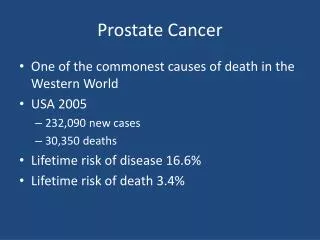
Prostate cancer • •By the age of 80, >80% of men have prostate cancer • •It is the second most common cancer in men, and the 4th most common cause of death for men in England and Wales. • •The tumours are adenomas and are usually located in the peripheral prostate. • •Spread is most commonly to bones. And prostate cancer is unusual in that it often causes an increased bone density- osteosclerosis (most cancers cause decreased bone density). • Aetiology • •Prostate cancer is rare before the age of 40, and prevalence increases with age. • •There is also a genetic factor – similar to that in breast cancer, so if someone in your family has the disease, then you are also more likely to have it. • •Average life-expectancy after diagnosis is approximately 5-10 years – the disease is very slow to progress. • Clinical features • •Lower Urinary tract obstruction • •Nocturia • •Poor flow of urine • •Symptoms of metastatic spread – particularly to the bones (weight loss and bony pain) • •Irregular hard prostate on DRE
Investigations • •PSA – this likely to be markedly raised in metastatic disease (>16µg/dl). You can also have a normal PSA and still have prostate cancer. • •DRE – it is NOT true that this elevates PSA levels • •Transrectal ultrasound of prostate (TRUS), often with biopsy. The ultrasound is useful because it gives a more accurate estimation of size than a DRE, and can also help stage any tumour present. You may also want to examine the upper renal tracts for signs of dilation. • •10-15% of TURP surgeries will uncover a cancer when it is only suspected to be BPH. • •Bone metastasis can be seen on X-ray as osteoscleoritc lesions. • Spread • •Bones – particularly the axial skeleton– these are detected with radionuclide bone scans • •Lymph nodes (obturator, internal iliac and presacral nodes) • •Bladder • •Rectum • •Seminal vesicles • •The scale for assessing the aggressiveness of the tumour is the Gleason score. This is a scale from 1-10, with 10 being the most aggressive cancers. • •For cancer spread, the scale of T1-4 is used.
Treatment • The condition is only curative when confined to the prostate. • •Finasteride – a 5α-reductase drug, It inhibits the production of dihydrotestosterone from testosterone and thus prevents growth of the prostate, however there is a high risk of sexual dysfunction. • •Watching and waitng – this is the management method proposed by most clinicians. The side effects of potential treatments are numerous and debilitating, and thus treatments are usually held back from as long as possible • •Radical prostatectomy – the is total removal of the prostate. It can be done ‘open’ or laparoscopically. The cure rate is 90% for tumours confined to the prostate. At 12 months, the incontinence rate is 7%, and impotence rate is 30% • •Radiotherapy – this, along with prostatectomy are the only curative treatments for prostate cancer. • ◦Brachytherapy – this is where radioactive ‘seeds’ are planted in the prostate. • •Androgen suppression – this is the main treatment for non-localised disease. About 80% of patients will show a sustained response to this treatment, but it can take 24-36 months for the response to appear. The quicker the PSA returns to the normal range, the better the prognosis. • ◦Luteinising hormone releasing hormone agonists – these stop the release of lutenising hormone, and thus the production of testosterone. • ◦Castration – many men refuse this treatment method.
Benign prostatic hypertrophy • This occurs most commonly in men over 60 • 24% of those ages 40-64, and 40% of those over 65 will have the condition. • Nearly All men will develop BPH if they live long enough. About ½ of all men will have macroscopic enlargement, and one half of these will have symptoms. • The prostate naturally grows throughout life – it grows in response to dihyrotestosterone – a breakdown product of testosterone. • Presentation • Frequency of urination (notably nocturia) is the most common early symptom • Hesitation in initiating urination. • Reduced force of the urinary stream • Post-void dribbling • Retention of urine resulting in overflow incontinence. • A benign prostate will always feel smooth • Size is relevant but is not always associated to the severity of the symptoms. • Investigations • PSA • PR (DRE – digital rectal examination) • Symptom score • Rectal ultrasound • Cystoscopy • Urine flow analysis
Management • Patients with mild symptoms should perhaps not be given treatment if they feel they can cope, due to the adverse effects many of the treatments. Sometimes symptoms after treatment may be worse than symptoms before treatment! • Patients with moderate symptoms should be treated with drugs: • α-blockers – alfuzosin, doxazosin, tamsulosin – these reduce smooth muscle contractions of the bladder and urethra – they generally reduce the muscle tension in these regions. This allows for easier passing of urine. • 5α- reductase – finasteride – these reduce the conversion of testosterone to dihyrotestosterone and thus help to shrink the size of the prostate. These take 4-6 months to have an effect. • Combination therapy – this is a combination of the two drugs above, and most patients who are on drugs for BPH are given this. • Patients with more severe symptoms should be considered for surgery. These symptoms may include renal damage, and upper UT dilation. • TURP – transurethral resection of the prostate – this is where part of the prostate is cut out via the urethra. It can be done under general or local anaesthetic. About 14% of patients will become impotent, and 20% of patients will need further surgery within 10 years. • In cases of acute retention, or retention with overflow, the immediate priority is to relieve discomfort and pain, and often catheterisation is a good idea. The urethral catheterization is not possible, then subra-pubic catheterization may be carried out. This involves sticking a tube through the abdominal wall directly into the bladder. It isoften done in cases where the patient needs to be catheterized for a prolonged period.
Testicular cancer • These are germ cell tumours. There are two main types (these account for 80% of all tumours) • Seminoma (dysgerminoma in women). These arise from the seminiferous tubules. They are a low-grade tumour and metastasis can occur via lymphatics and to the lungs. • Teratoma. A teratoma has a mixture of both mature and immature cells. initially they arise from germ cells, and often contain muscle, bone, fat and a variety of all sorts of other tissue. They are classified according to the degree of differentiation. As always, well differentiated tumours have the best prognosis. • Epidemiology • These account for 1-2% of all tumours • The incidence is very low, with prevalence being about 5 per 100 000 • They are the most common cancer in men ages 15-35 • Teratomas tend to occur in younger populations than seminomas • Presentation • Often they will be discovered incidentally as a firm lump on the testes. It may or may not be painful. • There may be some evidence of spread to para-aortic lymph node with associated back pain. • Some patients complain of a testicular ache.
Investigation • All suspicious testicular lumps should be examined by ultrasound. • There should be tests for serum tumour markers: • α-fetoprotein (AFP) • β-human chorionic gonadotrophin(HCG) • above markers tend to be increased more with more severe disease. • CT / MRI to check for distant metastasis (particularly in the lungs, liver, and retroperitoneally) • Treatment • Seminoma • They are both radio and chemo sensitive. Even with stage I disease, there is a 30% chance of recurrence. • Combination chemotherapy will cure 90% of those with metastatic disease. • 5-year survival is 90-95% • Teratoma • This is mostly treated with inguinal orchiectomy. One of the testicles will be removed via the inguinal route to minimise the risk of highly malignant cells spilling in the scrotum. The testicle and spermatic chord are removed to as far up as the inguinal ring. • Metastatic spread commonly involves the lungs and lymph nodes. • 80% of tumours will express AFP or HCG. • About 20% of men will be infertile at the time of diagnosis, but almost 100% of the rest will retain fertility and be able to father children. • 5-year survival is 60-95% depending on tumour stage and metastatic spread at the time of diagnosis.
Haematuria • Causes • Kidney • Trauma – mild to moderate trauma often causes this, but severe trauma may not. • Renal cell carcinoma – there may be loin pain, colic caused by a clot, an associated mass, hypertension, hypercalcemia, erythrocytosis (aka polycythemia –increased number of RBCs) • Calculus – severe loin / groin pain, associated infection • Pyelonephritis – (rare) • TCC – painless, intermittent haematuria • Ureter • Calculus – severe loin / groin pain, associated infection • Bladder • Calculus – sudden cessation of micturition, pain in perineum and penis • TCC – painless, intermittent haematuria, history of work in the rubber / dye industries. • Acute cystits – frequency, dysuria (pain / difficulty micturating), bacteriuria • Prostate • BPH – painless, haematuria, recurrent UTI, associated obstructive symptoms. • Carcinoma – rare cause of haematuria • Urethra • Trauma • Calculus – rare • Urethritis – rare • Investigations • FBC – to test for infection, and chronic blood loss • Clotting – to exclude an underlying bleeding cause • U+E – to assess renal function • MSU – to check for infection and parasites • Csytoscopy – if suspect a bladder cause • Autoimmune scan – if suspect glomerulonephritis • Intravenous Urography (IVU) / CT scan / ultrasound – if you suspect a renal cause
Renal cell cancer • This is an adenocarcinoma • Epidemiology • They are the most common renal carcinoma in adults. • They rarely present before the age of 40, and the average age of onset is 65-75 • Twice as common in men as women. • Aetiology • This is largely unknown, however some factors are suspected to precipitate this disease: • Irradiation • Exposure to oestrogens • Hypertension • Smoking • Exposure to cadmium • The disease is more commonly found in urban and industrial areas than in rural areas. • Incidence is 10 per 100 000 in males, and 5 per 100 000 in females.
Clinical Features • Often RCC is symptomless, until late stage. May be discovered incidentally. • The classic signs of this are: • Haematuria – 60%- this is caused by the spread of the tumour to the renal pelvis, which usually occurs very early on. From here it may spread to the renal vein and IVC. • Flank/loin pain (40%) • Palpable mass (25%) • These first three signs are the ‘classic’ ones, but actually only occur in 15% of patients. • Weight loss (30%) • Raised ESR • Polycythaemia (5%) • Hypertension (30%) – due to secretion of renin by the tumour • Anaemia (30%) – due to suppression of EPO by the tumour • Pyrexia of unknown origin (PUO) (20%) • Varicocoele (rare) – this occurs as a result of invasion of the left renal vein by the tumour, which may then affect drainage of blood from the testes. • However, many tumours are now discovered earlier as a result of incidental, or screening USS.
Treatment • The only way to treat these tumours is by surgical excision. • As long as you have at least half of one fully functioning kidney, then renal function will be adequate. • During total nephrectomy, the perirenal fat and fascia will also be removed. • Even in the presence of metastasis, nephrectomy is still recommended, as in many cases there is regression of the metastasis after removal of the kidney. In the case of a single metastasis, it is worthwhile to remove this secondary tumour as the metastasis is likely to be single due the relatively slow growing rate of renal cell carcinomas. • Advanced disease • For those with many metastasis, or invasion of the vena cava etc, then prognosis is generally very poor. Nephrectomy may still be performed to provide symptomatic relief, but most patients are unlikely to live longer than a year. • Medroxyprogesterone acetate may be useful in cases of metastatic disease. • Radiotherapy is only useful in treating bone mets. • Immunotherapy has been shown to be effective – treatment with interferon and interleukin-2 in patients with extensive disease is beneficial in 10-40% of cases, not necessarily ‘curing’ the disease, but it may prolong the patient’s life. In such cases, a nephrectomy will usually have been performed previously. Metastasis in the lungs are the most likely to respond to this sort of treatment. • Prognosis • 5 year survival: • Tumours confined to kidney – 60-70% • Lymph node spread – 15-35% • Metastatic disease – 5%
Stones • Renal stones are precipitates that form from urine due to a high concentration of that particular precipitate in the urine. The most common (85%) precipitate is calcium; particularly calcium oxalate, about 10% are uric acid, and 5% due to other precipitates. • They are likely to form where there is stasis, and they form more quickly once a nucleus has formed. • Epidemiology • More common in men • Occur in about 1/1000 individuals • By Age 70, 12% of men and 5% of women will have been affected • More common in elderly age groups – as they take years to form • Aetiology • Is generally multifactorial. It is believed that about 50% of cases are due to a hereditary disorder – hypercalcuriawhich results in increased urinary concentrations of calcium, despite normal serum ca2+ concentrations. • Hyperparathyroidism is also known to increase the risk due to its effects on calcium metabolism.
Presentation • Pain – a classical colicky loin pain. Patients often describe it as the worst pain they have ever felt! Patients will typically writhe around in pain and find it difficult to get comfortable. The ureters contract in a peristaltic manner, and move a ‘bolus’ of urine down from the pelvo-uteric junction (PUJ) down along the ureters. The pain felt in renal colic occurs every time the ureters contract and press onto the obstructing stone. The loin ain classically radiaties down and round (L1-L2 nerve routes). Nausea / VomitingHaematuriaSepsis • Fever (above 38, you become impaired, above 40 you may hallucinate). You increase your temperature to reduce the effectiveness of bacterial enzymes. • Dysuria (burning pain on micturation) • Tachycardia • Decreased blood pressure – can lead to septic shock. • Gram-negative bacteria are particularly dangerous • Differential diagnosis: • Pancreatitis • AAA • Gallstones • Musculoskeletal pain • Pyelonephritis
Investigations • Urinalysis – haematuria is common, but unless there is accompanying sepsis, urine will likely otherwise be normal. . • CTKUB • Management • Often conservative, especially if stone is less than 5mm. • Analgesia! • NSAIDs are useful as they aid with relaxation of ureteric smooth muscle. Diclofenac is usually the NSAID of choice a • Codeine/ morphine • Smooth muscle relaxants • Small smooth stones may pass themselvesStone may be passed depending on where it is, and urinary flow (which may be reduced due to renal failure)Mid-ureter – 60% chance of passing • Invasive management – if conservative is unsuccessful • Extracorporeal shock wave lithotripsy – using waves, break up the stone • Endoscopic – Ureteroscope • Endoscopic percutaneous • Endoscopic – laparoscopic very rare • Open operation. Very rare • Impacted stones will need intervention – this is where the contractions of the ureters will not move the stone, and this irritates with mucosa and lead to oedema, which then makes the stone even harder to pass! This can lead to stricture. • Good fluid intake is the best preventative measure for preventing stones. • Complications • Renal failure – any obstruction to the ureter will cause backflow and affect the GFR in the effected kidney. However, permanent damage is unlikely to result from hydronephrosis unless the obstruction is present for weeks at a time. • Sepsis – is more common than renal failure, but still generally unlikely.
TCC • This is a tumour of the bladder and urinary tract. It can occur anywhere along the urinary tract from the calyx, renal pelvis, ureter, bladder to the urethra. • Epidemiology • Uncommon before the age of 40, only 5% of cases present before the age of 60. • Male to female ratio 4:1 • Incidence is about 32 per 100 000 in men, and 10 per 100 000 in women. • Bladder tumours are by far the most common – 50x as common as tumours in any other region of the UT. • Peak age of presentation is 65-69 in men and 75-79 in women. • The incidence is declining thanks to the improvement of working conditions and supervision of workers is vulnerable occupations in the last 50 years. It is also expected that incidence will decline in the future in the West, due to changes in attitudes to smoking. • Aetiology • Smoking – it is thought that this accounts for 40% of cases of bladder cancerExposure to industrial chemical carcinogens; such as β-naphthylaminen and benzidine – these are found in: • Chemical, cable, rubber, leather, painting and dye industries. • Exposure to certain drugs; phenacetin, cyclophosphamideChronic inflammation
Clinical features • Painless haematuria is by far the most common presentation. In some cases, pain may occur due to clot retention. • There may be symptoms suggestive of a UTI, but urine will be negative for bacteria. • TCC of the kidney and ureter may give rise to flank pain (and haematuria) as a result of urinary tract obstruction. • Investigations • USS and CT • Analysis of urine for malignant cells. • Cystoscopy is also usually carried out, unless there is evidence that the malignancy is in the upper UT.
Treatment • Pelvic and ureteric tumours – are treated by nephroureterectomy. Radio and chemotherapies have been shown to be of little value. There should be follow up cystoscopy at regular intervals because 50% of these patients will develop subsequent bladder tumours. • Bladder tumours – treatment for these depends on the stage (described above): • pTa stage – these are treated by transurethral resection. Again, cystoscopy at regular intervals is necessary, as 70% will reccur • pT1 stage – these tumours have already shown their invasive potential – and they are treated in an unusual manner: • Intravesical BCG – this is the vaccine that is given foe TB. It stands for Bacillus Calmette-Guerin. It is given in bladder cancer as a form of immunotherapy. Even with this treatment, 50% of patients will develop invasive disease within 5 years. • pT2 stage and above – this is tumours that have invaded the muscle layers of the bladder or further. In patients under 70, treatment is with radical cystectomy. In patients over 70, treatment is with radiotherapy. Cystectomy carries a mortality risk of 2-4% which obviously rises with age. Both treatments have their downsides: • Prognosis • 5 year survival rates: • 80-90% for lesions not involving the bladder muscle • 5% for those with metastatic disease
Testicular Torsion • EPIDEMIOLOGY: Can occur at any age but most frequently among adolescents. It is rare >30 years of age. It occurs in about 1 in 160 males per year. • RISK FACTORS • Bell-clapper deformity (this is where the testicle has formed with no attachment to its surrounding scrotal walls and so is free floating within the tunica vaginalis – the serous sac surrounding the testicle) • An undescended testis • DIFFERENTIAL DIAGNOSIS • Epididymitis (main differential) • Epididymo-orchitis • Incarcerated inguinal hernia • CLINICAL PRESENTATION • Acute onset of diffuse pain – can be in the scrotum, groin, lower abdomen or the inguinal region. • Swollen testis • Testicular tenderness • Vomiting • INVESTIGATIONS • Doppler Ultrasound Scan can be done to look at the flow of testicular blood – this helps to rule out epididymitis where the flow will be present. In torsion, there will be absent blood flow. • Surgical exploration is mandatory unless torsion can be excluded. • TREATMENT • Surgical emergency– immediate intervention required to detort the testis. (also fix other side) • PROGNOSIS • If treated within 6 hours – 90% chance of survival of the testicle surviving; 12 hours – 50%; 24 hours – 10%; >24 hours – 0%.
Testicular/Scrotal lumps • Testicular cancer • Testicular torsion • Epididymo-orchitis • Epididymal cyst • Hydrocoele • Varicocoele • Inguinal hernia