Download
1 / 26
270 likes | 621 Views
VISUAL PATHWAY. BY PROF. SAEED ABUEL MAKAREM. Vision is the most highly developed and versatile of all the sensory modalities. The one which man are most dependent. The optic nerve and retina develop from the prosencephalon so, they are considered as an outgrowth of the brain itself.

E N D
VISUAL PATHWAY BY PROF. SAEED ABUEL MAKAREM
Vision is the most highly developed and versatile of all the sensory modalities. • The one which man are most dependent. • The optic nerve and retina develop from the prosencephalon so, they are considered as an outgrowth of the brainitself. Prof. saeed Makarem
The eyeball, or globe, is almost spherical in shape. • Near its posterior pole emerges the optic nerve. • The eyeball consist of three layers of tissue, the outermost, is tough, fibrous and protective. • Over most of the globe it forms an opaque white coat, the sclera,to which are attached the extraocular muscles. • Over the anterior pole of the globe it forms the transparentcorneathrough which light enters the eye. Prof. Saeed Makarem
Near the anterior margin of the sclera, two rings of smooth muscle extend into the lumen of the eyeball. • The anterior one is the iris,which has a central opening, called pupil,through which light passes to the posterior part of the eye. • The second one is the ciliary body, ciliary process, suspensory ligament and the lens. Prof. saeed Makarem
Some of the muscle fibers of the iris are arranged in a circularway while others are arranged radially. • Both fibers are under the control of the autonomic nervous system. • Circular fibersare innervated byparasympathetic neurones, which constrict the pupil and reduce the amount of light falling upon the retina. • Radial fibersare innervated by sympathetic neuronesto dilate the pupil. Prof. saeed Makarem
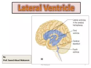
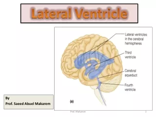
Vision starts with formation of an image on the photoreceptive retina. • Then the image discharged to the optic nerve. • Fibres of the optic nerve undergo hemidecussation in the optic chiasma. • Then it projects to the lateral geniculatenucleus of the thalamus through the optic tract. • Thalamo-cortical or (geniculo- calcarine)fibers in turn project to the primary visual cortex area17) through the retrolentiular part of the internal capsule to the occipital lobe where visual perception occurs. Prof. Saeed Makarem
Retinal photoreceptors are of two types, rods and cones. • Rods are about 20 times more than the cones. • These cells share many structural similarities. • Rodsare exquisitely sensitive to light. • They are particularly important for vision in dim light. • Conesare responsible for color vision,3 dimensions and, high visual acuity. • Rods lie more in the periphery towards the orraserrata, while cones are predominant at the macula and fovea centralis Prof. saeed Makarem
The first-order neurone, or bipolar cell,lies entirely within the retina. • While the axon of the second-order neurone, or ganglion cell, forms the optic nerve. In addition to photoreceptive cells, the retina contains both the first- and second-orderneurones of the central visual pathway. Prof. saeed Makarem
Information is transferred from photoreceptors to bipolar cells and then to ganglion cells. • The retina also contains interneurones known as horizontal cellsand amacrinecells. • These modulate transmissionbetween photoreceptors and bipolar cells and between bipolar cells & ganglion cells. Prof. saeed Makarem
The axons of ganglion cells assemble at the optic disc and pass into the optic nerve, which enters the cranial cavity through the optic canal. Prof. saeed Makarem
The two optic nerves converge to form the optic chiasma on the base of the brain. • The chiasma lies immediately rostralto the tuber cinereumof the hypothalamus and betweenthe terminating internal carotid arteries. Prof. Saeed Makarem
In the chiasma: • axons derived from the nasal portions of the two retinaedecussateand pass into the contralateral optic tract, • while those from the temporal hemiretinaremain ipsilateral. • Macular fibers pass to both optic tracts Prof. saeed Makarem
The optic tracts diverge away from the chiasma and pass round the cerebral peduncle to terminate mainly in the lateral geniculate nucleus(within the lateral geniculate body) of the thalamus. Prof. saeed Makarem
A relatively small number of fibers leave the optic nerve, before reaching the lateral geniculate nucleus,to terminate in the Pretectal area and the superior colliculus. Prof. saeed Makarem
These fibers are involved in mediation of the pupillary light reflex. Prof. Saeed Makarem
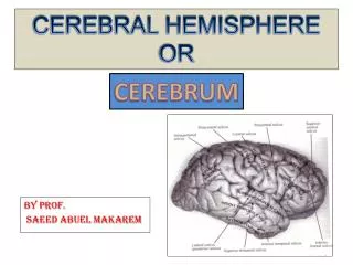
From lateral geniculate nucleus, third-order neurone (thalamocortical neurones) project through the retrolenticular part of the internal capsule and form the opticradiation,which terminates in the primary visual cortex of the occipital lobe. • The primary visual cortexis located predominantly on the medial surface of the hemisphere in the region above and below the calcarine sulcus. Prof. saeed Makarem
Surrounding this area, the rest of the occipital lobe constitutes the visual association cortex,( areas 18 &19). • It is concerned with interpretation of visual images, recognition, depth perception and colour vision. Prof. saeed Makarem
There is a precise point-to-point relationship between the retina and the visual cortex. • Because of the importance of the macula in vision, it is represented in both sides by disproportionately large volumes (relative to its size) in the lateral geniculate nucleus and the visual cortex. • Within the visual cortex the macula is represented most posteriorly, in the region of the occipital pole. Prof. saeed Makarem
The upper half of the visual field forms images upon the lower halves of the retina, the lower visual field upon the upper hemiretina. Prof. saeed Makarem
As thalamocortical fibersleave the lateral geniculate nucleus they pass around the lateral ventricle, those representing the lower part of the visual field coursing superiorly to terminate in the visual cortex above the calcarine sulcus. • Those which represent the upper part of the visual field sweep into the temporal lobe (Meyer's loop)before terminating below the calcarine sulcus. Prof. saeed Makarem
Visual field can be considered as comprising four quadrants(left/right, upper/lower) each projecting to its own quadrant of the primary visual cortex (left/right hemispheres, above/below the calcarine sulcus). • There is both lateral and vertical inversionin the projection of the visual field upon the visual cortex such that, for example, the upper left quadrant of the visual field is represented in the lower right quadrant of the visual cortex. Prof. saeed Makarem
Visual field deficits • Disease of the eyeball (cataract, intraocular haemorrhage, retinal detachment) & disease of the optic nerve (multiple sclerosis and optic nerve tumors) lead to loss of vision in the affected eye (monocular blindness). • Compression of the optic chiasm by an adjacent pituitary tumor leads to bitemporal hemianopia. • Vascular and neoplastic lesions of the optic tract, optic radiation or occipital cortex produce a contralateral homonymous hemianopia.
Retinitis Pigmentosa For more information, see http://www.emedicine.com/oph/topic704.htm • Retinitis pigmentosais an inheritedmetabolic disorder of the photoreceptor and retinal pigment epithelial cells. • It is due to mutation of a key protein in the retinal photoreceptors. • Which protein? • Rhodopsin. • There is progressive night blindness, peripheral visual field constriction and pigmentation of the retina visible on ophthalmoscopy. • Which type of photoreceptor is affected? • Rods.
Retinitis Pigmentosa • Can you name some of the genes whose mutation leads to retinitis Pigmentosa? • -RPGR (X-linked), RP1, chromosome -8 (http://www.nature.com/ng/journal/v22/n3/full/ng0799_248.html)
Retinitis Pigmentosa • Which part of the rods is affected first? • - The photoreceptor segment. • What happens next? • - The entire cells dies. • What you can do to help? • - Very little. • What drugs adversly affect RP? • Isoretinoin (Accutane) • Sildenafil (Viagra) • Vitamin E (high doses, >400 U/d) (http://content.nejm.org/cgi/content/abstract/323/19/1302 http://www.emedicine.com/oph/topic704.htm)