Download
1 / 20
200 likes | 231 Views
Get an in-depth look at the arrangement of muscles in the anterior and posterior compartments of the forearm, their origins, actions, and nerve supplies. Learn how injuries affect muscle function.

E N D
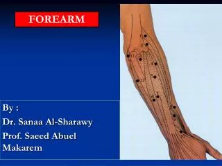
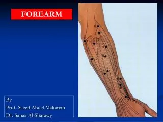
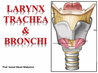
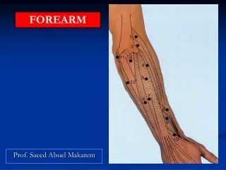
FOREARM Prof. Saeed Abuel Makarem
Objectives By the end of the lecture, you should be able to: Describe the arrangement of the muscles of the anterior and posterior compartments of the forearm. Describe the, origin, insertion, action nerve supply of each of these muscles. Describe the effect of injury of the muscle or its nerve supply.
The forearm extends from elbow to wrist. • It posses two bones radius laterally & Ulna medially. • The two bones are connected to each other by interosseous membrane. • This membrane allows movement of Pronation and Supination while the two bones are connected together. • Also it gives origin for the deep muscles.
Fascial Compartments of the Forearm The forearm is enclosed in a sheath of deep fascia, which is attached to the posterior border of the ulna . This fascial sheath, together with the interosseous membrane and the fibrous intermuscular septa, divides the forearm into anterior and posterior compartments, each having its own muscles, nerves, and blood supply.
Anterior compartment -FLEXOR GROUP These muscles: are (8) • They act on the elbow & wrist joints and the fingers. • They form fleshy masses in the proximal part and become tendinous in the distal part of the forearm. • They are arranged in three groups: I-Superficial: 4 • Pronator teres. • Flexor carpi radialis. • Palmaris longus. • Flexor carpi ulnaris. II-Intermediate: 1 • Flexor digitorum superficialis. III- Deep: 3 • Flexor digitorum profundus. • Flexor pollicis longus. • Pronator quadratus.
Superficial Flexors • They arise - more or less- from the common flexor origin (front of medial epicondyle). • All are supplied by median nerve exceptone, flexor carpi ulnaris, (FCU) which is supplied by the ulnar n. • All cross the wrist joint except one, pronator teres, (PT).
Flexor Carpi Radialis • Insertion: Base of 2nd metacarpal bone • Action: Flexion & abduction of the wrist. • Palmaris Longus • Insertion: into the flexor retinaculum & the palmer aponeurosis. • Action: Flexes hand & tightens the palmer aponeurosis. • Pronator teres Insertion: middle of lat. surface of radius • Action: pronation & flexion of forearm. May Be Absent
Flexor Digitorum Superficialis • Insertion: • Base of middle • phalanges of • the medial 4 • fingers. • Action: • Flexes middle • and proximal • phalanges of the • medial 4 fingers, • Flexion of the hand (wrist). • Flexor Carpi Ulnaris • Insertion: • Pisiform, • Hook of hamate • 5th metacarpal bone. • Action: • Flexion and adduction of the hand (wrist).
Origin of the Deep Flexors (3 muscles) • Front of radius: Flexor pollicis longus. • Front of ulna: Flexor Digitorum profundus. • Front of lower 4th of ulna . Pronator Quadratus.
Pronator Quadratus • Insertion: distal fourth of anterior surface of radius. • Action: pronates the forearm (primover). • Hold the 2 bones together . • Flexor Pollicis Longus • Insertion: Base of distal phalanx of thumb. • Action: flexes all joints of the thumb. • Flexor Digitorum Profundus • Insertion: bases of distal phalanges of the medial four digits. • Action: Flexes distal phalanges of medial four digits.
Nerve supply of the deep flexors All are supplied by the anterior interosseous nerve (branch of the median nerve), Except the medial half of the flexor digitorum profundus which is supplied by the ulnar nerve.
Supination and pronation It occurs in the superior and inferior radioulnar joints; (Pivot Uniaxial Synovial Joint.) Muscles produce supination Biceps brachii. Supinator. Muscles produce pronation Pronator teres. Pronator quadratus. NB. Brachioradialis put the forearm in midprone-supine position.
Posterior compartment: 3 groups Superficial group 5 muscles: • Extensor carpi radialis brevis. • Extensor digitorum. وبس • Extensor digiti minimi. • Extensor carpi ulnaris. • Anconeus. Origin: Common Extensor Origin. (front of the lateral epicondyle). Deep group: 5 (3 to thumb+ 1 to index + Supinator). • Abductor pollicis longus. • Extensor pollicis brevis. • Extensor pollicis longus. • Extensor indices. • Supinator. Lateral group 2 muscles: • Brachioradialis. • Extensor carpi radialis longus. (The 2 muscles originate from the lateral supracondylar ridge).
Posterior compartment • I- Superficial group: • 7 muscles ( from lateral to medial) • Brachioradialis, (BR). • Extensor carpi radialis longus, (ECRL). • Extensor carpi radialis brevis, (ECRB). • Extensor digitorum, (ED).وبس • Extensor digiti minimi, (EDM). • Extensor carpi ulnaris, (ECU). • Anconeus. (An).
Superficial Extensors • All arises from the commonextensor origin, (front of lateral epicondyle) of the humerus, EXCEPT, 3 (BR, EXRL & anconeus). • All cross the wrist EXCEPT, 2, (brachioradialis & anconeus). • All supplied by deep branch of radial nerve, EXCEPTABE • A, Anconeus. • B, Brachioradialis. • E, Extensor carpi radialis longus. • These 3 muscles are supplied by the radial nerve itself.
Extensor Carpi Radialis longus • Origin: • Lateral supracondylar ridge of humerus. • Insertion: • Posterior surface of base of second metacarpal bone. • Action: • Extends and abducts the hand at wrist joint. • Brachioradialis • Origin: • Lateral supracondylar ridge of humerus. • Insertion: • Base of styloid process of radius. • Action: • Flexes forearm; (elbow). • Rotates forearm to the midprone position.
INSERTION Extensor carpi radialis brevis: base of 3rd metacarpal bone. Extensor digitorum: Extensor expansion of the medial 4 fingers. Extensor digiti minimi: Extensor expansion of the little finger. Extensor carpi ulnaris: Base of the 5th metacarpal bone.
II- Deep group: 5 muscles, (3 to thumb+1 index+ Supinator). 1- Abductor pollicis longus, (APL). 2- Extensor pollicis brevis, (EPB). 3- Extensor pollicis longus, (EPL). 4- Extensor indicis (EI). 5- Supinator. All back muscles of forearm are supplied by posterior interosseous nerve except , ABE by Radial nerve.
Dorsal Extensor Expansion It is formed by the union of the tendons of: Extensor digitorum, Extensor indicis, extensor digiti minimi, palmar & dorsal interossei and lumbricals muscles. All these tendons unite to form one tendon which divides into 3 slips, a median one attached to middle phalanges and 2 lateral attached to the terminal phalanges.