Monocoli-3d
C. A. GF. Monocoli-3d. GF. Monocoli-3d. Monocoli-7d. Monocoli-21d. CV. GF. Monocoli-3d. Monocoli-21d. D. B. Monocoli-7d. Monocoli-21d. GF. Monocoli-3d. Monocoli-7d. Monocoli-21d. CV.

Monocoli-3d
E N D
Presentation Transcript
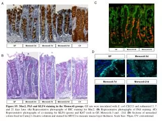
C A GF Monocoli-3d GF Monocoli-3d Monocoli-7d Monocoli-21d CV GF Monocoli-3d Monocoli-21d D B Monocoli-7d Monocoli-21d GF Monocoli-3d Monocoli-7d Monocoli-21d CV Figure S5: Muc2, PAS and KLF4 staining in the Monocoli groups. GF rats were inoculated with E. coli CEC15 and euthanized 3, 7 and 21 days later. (A) Representative photographs of IHC staining for Muc2. (B) Representative photographs of PAS staining. (C) Representative photographs of co-staining for KLF4 (green) and Ki67 (red) in GF, Monocoli-3 and -21d. (D) Sections of unwashed colons fixed in Carnoy’s fixative solution and stained for MUC2 to measure mucus layer thickness. Scale bars: 50µm. CV: conventional.