Case 10
Case 10. New Frontiers in Pathology, 2008 William G. Finn, M.D. University of Michigan Ann Arbor, Michigan. Case Presentation.

Case 10
E N D
Presentation Transcript
Case 10 New Frontiers in Pathology, 2008 William G. Finn, M.D. University of Michigan Ann Arbor, Michigan
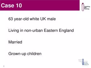
Case Presentation • A 79 year old man presented with cervical lymphadenopathy and a lesion at the base of the tongue. The tongue lesion was biopsied. He was subsequently admitted to the hospital with progressive dyspnea, cough, and pulmonary infiltrates on chest X-ray. Upper endoscopy had revealed “prominent gastric folds” and subsequent abdominal CT scan showed prominent splenomegaly, periaortic lymphadenopathy, and “stranding” of mesentery. He underwent a cervical lymph node biopsy 9 days after the tongue biopsy.
TCR-gamma PCR, cervical lymph node v1-8 v 9 -globin v 10-11
IgH PCR, cervical lymph node FR1 FR2 -globin FR3
Summary • Diffuse CD20 positive infiltrate of tongue, diffusely EBER + • T-cell infiltrate in lymph node • Arborizing vasculature • Disrupted dendritic meshwork • EBER+ individually scattered immunoblasts • Lymph node shows both T and B clonal gene rearrangements
Diagnosis Diffuse large B-cell lymphoma (EBV+) arising in the setting of angioimmunoblastic T-cell lymphoma
Pathogenesis of AILTL • Neoplasm of germinal center T-cells • CD4+ • CXCL13+ • CD10+ • Bcl-6+ • Systemic signs/ symptoms • Hypergammaglobulinemia, rash, fever, etc
Allen et al: Immunity 2007; 27:190
Pathogenesis (?EBV cause or effect) Immune dysregulation AILT EBV DLBCL
Summary – AILT lymphoma • A distinct clinico-pathologic entity • Lymphoma of T helper cells that normally reside in the germinal center and function in antigen-driven B cell development • Typically harbors EBV positive B cells • Occasionally the EBV+ B cells proliferate • In extreme cases the B cell proliferation progresses to frank DLBCL