Download
1 / 63
630 likes | 907 Views
November 29 th Therapeutic approaches in AD: -Beta and gamma secretase inhibitors -Abeta immunotherapy -Tau -Tau immunotherapy -Ongoing treatments in AD. Model for g -secretase complex and its interaction to the substrates.

E N D
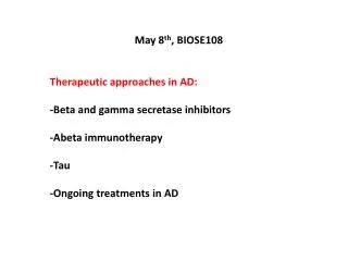
November 29th Therapeutic approaches in AD: -Beta and gamma secretase inhibitors -Abeta immunotherapy -Tau -Tau immunotherapy -Ongoing treatments in AD
Model for g-secretase complex and its interaction to the substrates The bars represent the transmembrane domains of the the proteins constituting the g-secretase complex
b- and g-secretase : possible therapeutic targets in AD?
Could BACE be considered as a therapeutic target for AD? In favor: 1- BACE is the primary b-secretase aspartyl protease that cleaves APP generating b-amyloid species (secondary role of BACE2) 2-BACE KO mice DO NOT have amyloidogenic processing of APP Against: 1-BACE+/- heterozygous mice do not show altered production of b-amyloid and APP C-terminal fragments 2-BACE cleaves a number of other substrates, including *Amyloid Precursor Like Proteins APLP1 and APLP2 *Low-density lipoprotein receptor (LDLR)-related protein LRP Thus, targeting BACE for the treatment of AD would result in loss of BACE activity towards the other substrates, with possible consequent implications on their physiological function.
APP processing and Ab 42 production are reduced in BACE knockdown mice Singer et al.,
Ab load and plaque size are reduced in BACE knockdown mice Singer et al.,
BACE KO mice show hypomyelination of hippocampal neurons… Hu et al.,
…and of the optic and sciatic nerves Hu et al.,
BACE KO- mediated hypomyelination is due to reduced activation of neuregulin Hu et al.,
BACE inhibition reduces or delays myelination in vitro BACE inhibitor ADAM inhibitor
Thus reducing BACE levels (or activity) may lead to neurodegeneration as side effect
Non-peptidic BACE inhibitor reduces Abeta levels in AD mice and Dogs
Non-peptidic BACE inhibitor reduces Abeta levels in healthy volunteers in a dose-dependent fashion
Non-peptidic BACE inhibitor reduces Ab levels in both plasma and CSF
BACE inhibitor causes cytosolic accumulation of granular material This effect is NOT associated to its role as beta-secretase inhibitor Vehicle BACE inhibitor Rat retinal epithelium BACEKO retinal epithelium
APP intrabodies, to affect APP transport and BACE-mediated amyloidogenic processing sFvb1 sFvb1-KDEL
APP intrabody carrying a KDEL motif for ER retention impairs APP maturation
g-secretase cleaves different substrates Bart De Strooper What about targeting g-secretase for the treatment of AD? Some of these substrates have crucial activity in regulating cell fate decision. In this respect, targeting g-secretase for the treatment of AD is not an easy task, as blocking g-secretase activity would have consequences on the physiological functions of the other protein substrates.
g-secretase, in particular PS1 and PS2 are not an easy therapeutic target for the treatment of AD Could Nicastrin be a good target?
The mechanism by which nicastrin selectively recognizes the substrates Nicastrin recognizes specific sequences in the N-terminal portion of C99 (and possibly of C83) and Notch C-terminal fragment. Shah et al.,
Mechanism by which Nicastrin participates in the “Regulated Intracellular Proteolysis” RIP BACE b-secretase Steps: 1-The substrates gets in close proximity with the g-secretase complex (i.e. after internalization from the plasma membrane). 2-Nicastrin specifically recognizes the substrate, and binds to it. 3-Presenilin (PS1) cuts the substrate within the exposed sequence (in the case of APP will be the Ab sequence). Shah et al.,
Chemical blocking of the N-terminal portion of the substrates will regulate nicastrin capability to recognize and to bind to it. Implications for the treatment for AD Will NOT bind to Nicastrin Shah et al., Will bind to Nicastrin
How to target Ab oligomers? Using molecules that interfere with the structure of the oligomer and break it up to single Ab monomers. Advantages of this therapeutic approach would be: 1-decreased accumulation of Ab oligomers, thus reduced formation of b-amyloid plaques 2-the single Ab monomers have higher chances to be removed by the action of clearing enzymes like neprilysin or Insulin Degrading Enzyme IDE 3-both intracellular and extracellular formed Ab oligomers would be targeted and disrupted.
New approaches for future therapeutic intervention in AD 1-molecules that disrupt the structure of the Ab oligomers 2-use of selected b- or g-secretase inhibitors. 3-used of vaccines, that remove the already deposited plaque
Decreased Ab plaque burden in animals overexpressing Insulin Degrading Enzyme (IDE) and Neprilysin Leissring et al.,
Ab immunization: clearing Ab plaques Hyman and Growdon, Nature Medicine 2006 Vol 12 (7): 755
Model of response to Ab vaccine Greenberg et al.,
Abeta immuno-therapy reduces the plaque load in AD animal model
Before immunization, AD patients show plaques with dystrophic neurites (a) and tau staining (b) Nicoll et al.,
Areas devoid of Ab plaques (c) do show NFTs but not dystrophic neurites (d) Nicoll et al.,
Areas devoid of plaques show punctate Ab staining Nicoll et al.,
Meningoencephalites in AD patients treated with Ab42 vaccine Lymphocytes in the leptomeningis T-lymphocytes Nicoll et al.,
Macrophages infiltrates the cerebral white matter in patients treated with Ab42 vaccine Infiltration of cerebral white matter by macrophages Vacuolation and refraction in myelinated fibers in the white matter Nicoll et al.,
Deposition of fibrillar proteinacious material in Alzheimer’s disease Alzheimer’s disease: characterized by extracellular depositions, the b-amyloid plaque, and intracellular depositions, the Neurofibrillary Tangles(NFT) comprised of Paired Helical Filaments (PHF), aggregates of hyperphosphorylated protein tau. Bossy-Wetzel E, et al., Nat Med. 2004 Jul;10 Suppl:S2-9. Review.
Origin of PHF and NFT 1-NFT are composed of paired helical filaments (PHF), aggregates of phosphorylated protein tau that form when levels of phosphorylated tau are elevated in the cell. 2-Tau is a microtubule-associated protein that regulates cytoskeleton structure. When highly phosphorylated, tau is sequestered into PHF, and causes disruption of microtubules, that ultimately leads to cell death. 3-Phosphorylation of tau by protein kinases such as the neuron-specific cyclin dependent kinase 5 (cdk5) precedes the formation of PHF that cause neurodegeneration. 4-Importantly, the formation of PHF and NFT is a hallmark in AD and many different neurodegenerative diseases, which together are called tauopathies.
Tau binds to microtubules regulating their connections with other cytoskeletal components such as neurofilaments This function is regulated by phospho/dephospho state of tau www.emdbiosciences.com
Tau hyperphosphorylation is not specific of AD, but occurs in other NADD