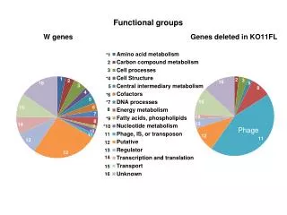
Phage
Phage. Ethan Callies. Purpose. To collect, recreate and analyze phage from the environment To sequence DNA from phage. Background. Virus that replicates using bacteria Ernest Hankin , 1896 India, something in water cured Cholera Frederick Twort , 1915


Phage
E N D
Presentation Transcript
Phage Ethan Callies
Purpose • To collect, recreate and analyze phage from the environment • To sequence DNA from phage
Background • Virus that replicates using bacteria • Ernest Hankin, 1896 • India, something in water cured Cholera • Frederick Twort, 1915 • Discovered small agent that killed bacteria • Work interrupted by WWI • Félix d'Herelle • September 3, 1917 discovered “an invisible, antagonistic microbe
Phage Therapy • Phage used as antibacterial agents • Began in 1920’s • Today is most common for fighting Staphylococcus, Streptococcus, and other infections
My Phage • Collected from base of tree in River Falls, WI • Moist soil conditions • About 44mm below surface • Lola13
Enrichment of Environmental Samples • Did this to increase the chance I will get a phage from my sample
Phage Titer/Streak Plate • Titer • Determine concentration of Plaque forming units (pfu) • Streak Plate • Help purify single phage population from mixed populations
Phage Purification • Isolate enough pure phage sample to create Lysate or MTL (Medium Titer Lysate)
Phage Lysate • Used to create large batch of pure phage called HTL (High Titer Lysate)
Web Plates • Obtain HTL with high enough phage concentration to isolate DNA
Isolate/Purify Genomic DNA • Want to isolate this in high enough amounts for restriction analysis/sequencing
Gel Electrophoresis • To compare phage DNA sample to a known DNA sample
Digest Phage DNA • See how Phage DNA compares to known samples when digested by different enzymes
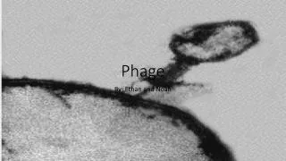
Electron Microscopy • Observe individual phage using electron microscopy
Results (After 3 enrichments) • 3 different morphologies developed • Created streak plate for each • Smaller and bigger morphologies purified very well • Titrated each out separately with 4 dilutions
Results (after discovering morphologies) • Smaller morphology to 2nd dilution • Bigger had plaques to 4th dilution Small titer: 4.6x10^-4 pfu/mL Bigger titer: 5.4x10^-6 pfu/mL • Repeat purification
Results (after repeating purification) • Smaller morphology disappeared • Bigger morphology to all plates (8 in 10^-2, 2 in 10^-3 and 1 in 10^-4)
Spot test to determine web plate Countable plate to determine titer Titer: 2.8x10^-4 pfu/mL
Web Plate • Plate webbed very well • Small morphology appeared again • Set up 10 web plates
Harvest HTL/Titer/Quanitification • All 10 plates completely plaqued out • Titer came out to be 2.75x10^-9 pfu/mL • Spectrophotometer read .119 mg/mL
EM Picture • Tails are approximately 70 nm • Heads are approximately 30 nm
Conclusion • Many morphologies appeared throughout purifications. While this could be because of contamination, I think it was because of something biological. This could be improved a bit by making things a bit more “sterile” while going through the purification process. • What caused the different sized plaques to show up? Contamination or biology?