Download
1 / 23
240 likes | 496 Views
The Secretory Pathway Becky Dutch Molecular and Cellular Biochemistry 1. ER - translation 2. ER- protein modifications 3. Discussion Section 4. Golgi apparatus 5. Vesicular transport. Lecture 3: The Golgi apparatus. Reading: Alberts Chapter 13

E N D
The Secretory Pathway Becky Dutch Molecular and Cellular Biochemistry 1. ER - translation 2. ER- protein modifications 3. Discussion Section 4. Golgi apparatus 5. Vesicular transport
Lecture 3: The Golgi apparatus Reading: Alberts Chapter 13 Lodish Sections 17.7 and 17.8
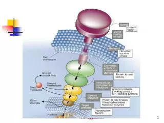
Modification and Sorting in the Golgi: 1. Addition, Processing of carbohydrates - ER, Golgi 2. Specific Proteolytic cleavages - Golgi 3. Sorting to proper destinations . Lodish, Fig 17-13
Golgi Apparatus: collection of flattened, membrane-bound cisternae. Alberts 13-3
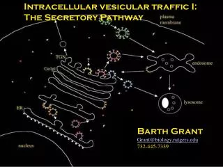
Proteins from ER enter at cis face, go through different compartments - then leave TGN for their final destinations. Alberts 13-4
Cisternal Progression or vesicular transport? -How do proteins move from cis to medial to trans? -Originally thought to be by transport vesicles (COP1). Alberts 13-14
Cisternal Progression/Maturation Evidence - Biosynthetic aggregates (collagen precursors) too large for vesicles traverse the Golgi stack. No evidence for a megavesicle. Rate of aggregate movement much slower than for normal proteins. Model - cisternae “progress” or mature. Cis Golgi becomes medial becomes trans. Golgi enzymes are moved back by retrograde transport.
Vesicular Transport in Golgi Evidence: Two distinct forms of COP1 vesicles - those with KDEL receptor and little cargo, and those with lots of cargo, no KDEL receptor. What is this second population doing? Also, transport for many proteins is faster than seen for aggregates. Model: COP1 vesicles mediate transport from cis to medial to trans.
Cisternal Progression and vesicular transport Model: Both may function in transport “Slow Track” - cisternal progression “Fast Track” - COP1 vesicles Pelham and Rothman, Cell 102: 713-719. Alberts 13-14
Processing glycoproteins in Golgi Cis, medial and trans compartments have different enzymes High mannose form - reactions 1 or 2 do not occur - Man8(GlcNAc2) or Man5(GlcNac2) - do not get further additions in Golgi Complex carbohydrates - additions in the Golgi Lodish 17-38
Endo H digestion distinguishes complex from high mannose Alberts 13-11
How do nucleotide sugars get into the Golgi? Antiporters located in the Golgi membrane One-for-one exchange keeps the concentration of sugar nucleotides constant Lodish 17-33
Targeting proteins to the lysosome Addition of P to Mannose 6 is needed for recognition by the Mannose - 6 - P receptor Signal sequence binds recognition site, catalytic site is distinct Lodish 17-39
Mannose - 6- phosphate pathway Lysosomal enzymes phosphorylated Bind M6P receptor in TGN Directs incorporation into clathrin-coated vesicle Uncoating, fusion with late endosome Low pH of endosome results in receptor release - P removed Receptor recycles, enzyme goes to lysosome Lodish 17-40
Disorders in lysosomal enzyme sorting Inclusion cell disease (I cell disease) - rare disorder in which almost all hydrolytic enzymes are missing from lysosome I cell disease - single gene, recessive defect Hydolases found in blood instead Defective or missing GlcNAc-phospho- transferase No P, no binding M6P receptors Some cell types (heptacytes) still sort to lysosome - must be an M6P independent pathway Alberts 13-23
Retention of resident Golgi Proteins All made in ER - carbohydrate modifying enzymes have similar structure: short N-terminal domain that faces cytosol (so Type II), single TM a helix, large C-terminal domain facing Golgi lumen Membrane spanning domain necessary and sufficient for retention in the Golgi - mechanism of retention not known. Not one single sequence of TM. Lodish 17-21
Sorting into Vesicles TGN to plasma membrane - two major types of vesicles: secretory vesicles for regulated secretion and transport vesicles for constitutive secretion How do proteins get sorted to the correct vesicle? Common mechanism seems to function for many regulated secretory proteins (ACTH, insulin, trypsinogen) but don’t share common sequence. Hypothesis: selective protein aggregation. Many mammalian cells have secretory vesicles with chromogranin B and secrotogranin II - form aggregates in TGN conditions (pH 6.5, 1mM Ca)
Proteolytic processing in the TGN Most secretory proteins, some plasma membrane proteins - made as proproteins - must be proteolytically cleaved to be active. Examples include insulin, glucagon, HIV env Some of these cleavage events occur in secretory vesicles as they leave TGN Insulin - only mature vesicles show cleaved form Ab to proinsulin Ab to insulin Lodish 17-41
Some proteins have single cut site Furin - endoprotease. In all mammalian cells. In the Kex2 family of endoproteases. Lodish 17-42a
Some secreted proteins undergo multiple cleavages Proinsulin - regulated secretion. PC2 and PC3 endoproteases - also in Kex2 family. Only found in cells with regulated secretion. Localized to secretory vesicles. Three cleavage events needed to form insulin. Lodish 17-42b
Apical/Basolateral Sorting Polarized epithelial cells - plasma membrane has two domains: apical and basolateral. Separated by tight junctions. Proteins sorted to one or other membrane One way: sorting in TGN - distinct vesicles go to the two surfaces Examples Influenza HA - apical VSV G - basolateral GPI-linked - apical (in endothelial; basolateral in thyroid) No unique sequence for targetting Lodish 17-43
Apical/Basolateral Sorting II Hepatocytes - another sorting mechanism. All proteins targetted to basolateral membrane Both types endocytosed Basolateral recycled to basolateral membrane Apical move to apical - process called transcytosis Lodish 17-43
Next lecture: Vesicular Transport . Lodish, Fig 17-13