Download
1 / 25
260 likes | 323 Views
Learn about the comprehensive management of Spinal Muscular Atrophy (SMA) Type 1, including its history, examination techniques, investigative procedures, treatment options, and prognosis. Understand the clinical manifestations and diagnostic features associated with SMA Type 1.

E N D
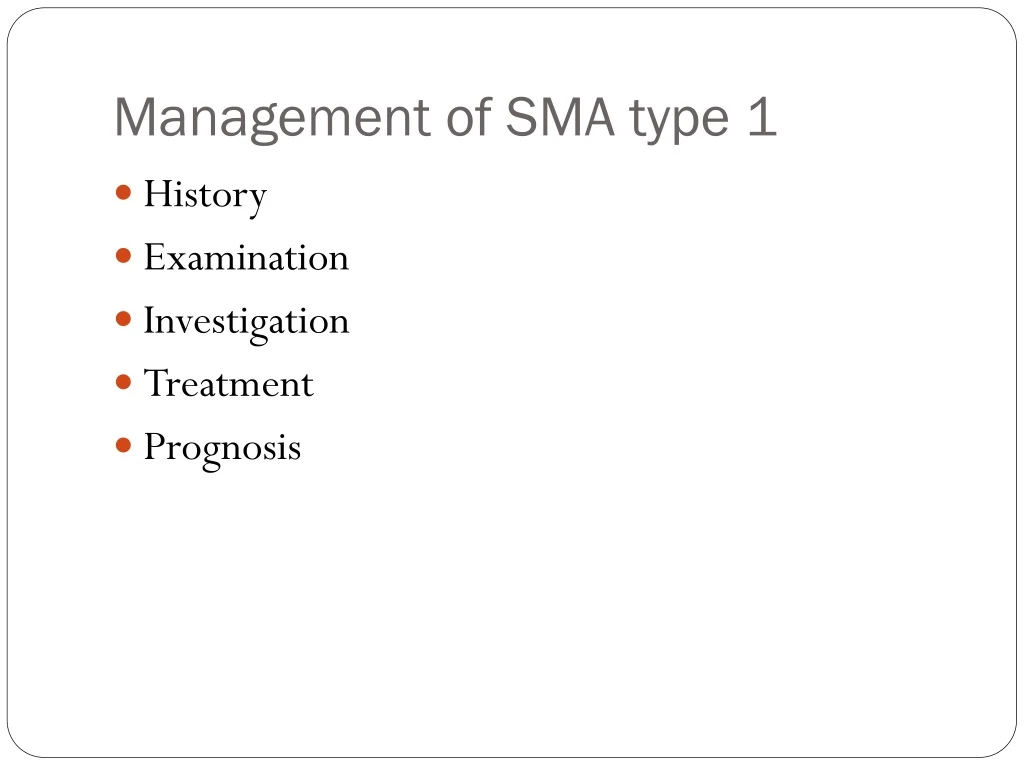
Management of SMA type 1 • History • Examination • Investigation • Treatment • Prognosis
History & Examination • Prenatal Hx – decreased fetal movement • Neonatal Hx – asphyxia, kernicterus, IVH etc • Hx of suspected aetiology • Muscle bulk • Tone • Strength • Distribution of weakness and muscle wasting • Proximal - Myopathy • Distal - Neuropathy • Myotonia is specific for a few myopathies
Tendon stretch reflexes • Lost - in neuropathies and in motor neuron diseases • Diminished but preserved - in myopathy • Fasciculations of muscle – tongue • Present • Absent • Sensory abnormalities - neuropathy • Involvement of the face, tongue, palate, and extra ocular muscles
Muscle pain or myalgias • Present - Acute disease of either myopathic or neurogenic origin. • Acute dermatomyositis • Acute polyneuropathy (Guillain-Barré syndrome) etc • Several metabolic diseases of muscle and in ischemic myopathy • Absent • Muscular dystrophies and spinal muscular atrophies • Contractures of muscles – at birth or developing later • Myopathic and neurogenic diseases.
Descent of testes • Undescended - Gubernaculum is weakened • Spinal muscular atrophy • Myotonic muscular dystrophy • Many congenital myopathies. • Shape of thorax • funnel shape – IUGR • Infantile spinal muscular atrophy • Myotubular myopathy • Neonatal myotonic dystrophy etc
Spinal Muscular Atrophies • Degenerative diseases of motor neurons • 2nd most common neuromuscular disease • Most cases are inherited as an autosomal recessive trait. Incidence of SMA is 10–15 per 100,000 live births • No racial predilection • Genetic locus for all 3 of the common forms of SMA is on chromosome 5
Onset • In fetal life • Continue to be progressive in infancy and childhood • Upper motor neurons remain normal. • Classification based on clinical manifestations • Age at onset • Severity of weakness • Clinical course
SMA type 1 - Werdnig-Hoffmann Dx • Severe infantile form • 25% of cases • SMA type 2 - Dubowitz disease • Late infantile and more slowly progressive form • 50% of cases • SMA type 3 - Kugelberg-Welander disease • More chronic or juvenile form • 25% of cases.
SMA type O • Severe fetal form that is usually lethal in the perinatal period • Rare and accounts for <1%. • A variant of SMA - Fazio-Londe disease • Motor neuron degeneration more in the brainstem than the spinal cord. • Progressive bulbar palsy • Some patients are transitional between types 1 and 2 or between types 2 and 3 in terms of clinical function
SMA type 1 – Werdnig-Hoffmann disease • Johann Hoffmann and Guido Werdnig • A genetic disorder • Inheritance is autosomal recessive • Results from destruction & degeneration of motor neurones in the spinal cord and the brain stem in utero following prolongation of the normal process of programmed cell death • It is the most severe form of SMA • Starts at birth or within 6 mo of age • Incidence is 0.4 per 1000 births.
Clinical Features • Reduced foetal movements in utero • Infant is severely hypotonic at birth with a frog-like posture • Triad of symptoms • Severe hypotonia • Absent reflexes • Fasciculation - tongue & fingers • Severe weakness in a proximal > distal distribution is seen • Thin muscle mass
Absent tendon stretch reflexes • Involvement of the tongue, face, and jaw muscles • Tongue fasciculation – diagnostic • Respiratory muscles are weak, and intercostal muscles are usually affected more than diaphragm, leading to paradoxical abdominal movement and pectus excavatum • Difficulty with swallowing & accumulation of secretions in the mouth because of weak masticatory and pharyngeal muscles • Inability to feed • Aspiration pneumonitis
Congenital contractures • Simple clubfoot • Generalized arthrogryposis • 10% of severely involved neonates • Unable to overcome gravity • Neck control grossly delayed • Weak cry • Unusual delay in achieving developmental milestones • Infants usually not strong enough to sit or sit late
No sphincteric or sensory impairment • Extra-ocular muscles are unaffected • Facial muscles are not weak • The alert state is not affected • Mental development is normal • Normal IQ • Children often appear brighter than their normal peers • Myalgias are not a feature of SMA. • The heart is not involved in SMA. • About 70% die either in early infancy or before the age of 2 yrs
Investigation • Molecular Genetic Studies • Genetic marker in blood for the SMN gene • Simplest, Highly sensitive and specific • By DNA probes in blood samples or in muscle biopsy or chorionic villi tissues • Serum CK level • Isoenzymes • MM for skeletal muscle • MB for cardiac muscle • BB for brain • Normal • Mild elevation
Nerve conduction velocity (NCV) • 80% of the total nerve fibers must be involved before slowing in conduction is detected. • Motor • Normal - usually • Mild slowing in terminal stages of the disease • Sensory may be slowed • Electromyography (EMG) • Shows fasciculations and fibrillations • Muscle Biopsy • Classic finding is grouped atrophy
Treatment • No medical treatment delays the progression • Supportive therapy • Orthopedic care - scoliosis and joint contractures • Mild physiotherapy • Spine fusion • Respiratory care • Non-invasive ventilation • Tracheostomy • Nutritional care • Tube feeding
Mechanical aids for assisting the child to eat and to be as functionally independent as possible • Respiratory depression is the most serious complication and the most common cause of mortality. • Aggressive and early respiratory toilet and treatment is required. • Most Type 1 patients will have severe respiratory compromise and may eventually mechanical ventilation
Prognosis • Poor • Many Type 1 and Type 2 patients do not survive childhood without mechanical ventilation • Do not survive beyond 3yrs • 7months to 7yrs for milder forms
References • Asindi AA. Muscular dystrophies and the floppy infant. In: Azubuike JC, Nkanginieme KEO, ed(s). Paediatrics and child health in the tropics. Owerri: African Educational services; 2004. p. 520-4 • Sarnat HB. Neuromuscular Disorders. In: Kleigman RM, Behrman RE, Jenson HB, Stanton BF, ed(s). Nelson textbook of Pediatrics. Philadelphia: WB Saunder Company; 2008. p. • Bodensteiner JB. The evaluation of hypotonic infant. Semin Paediatr Neurol 2008; 15:10-20