Download
1 / 74
740 likes | 816 Views
Explore the intricate parts of long bones, bone development, function, fractures, and types of fractures. Learn about cranial bones, bone tissue homeostasis, and the role of facial bones in supporting the face structure.

E N D
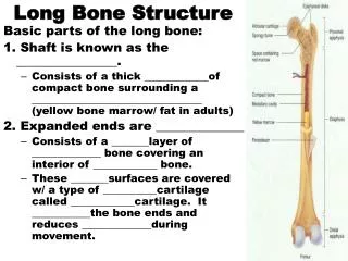
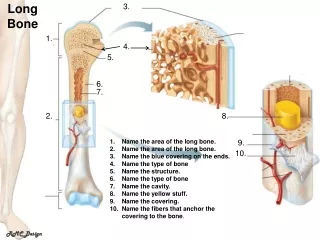
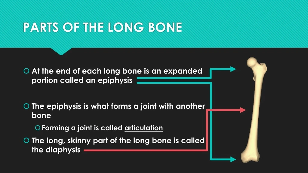
PARTS OF THE LONG BONE • At the end of each long bone is an expanded portion called an epiphysis • The epiphysis is what forms a joint with another bone • Forming a joint is called articulation • The long, skinny part of the long bone is called the diaphysis
PARTS OF THE LONG BONE • The outer surface of the articulating portion of the epiphysis is coated with articular cartilage • A tough covering of dense connective tissue called the periosteum completely encloses the bone except for the cartilage on the bone’s ends • The periosteum also helps form and repair bone tissue • A bone’s shape makes the bone’s function possible!
PARTS OF THE LONG BONE • The walls of the diaphysis are mainly composed of tightly packed tissue called compact bone • The walls of the emphysis are composed of spongy bone • Compact bone in the diaphysis form a tube with a hollow chamber called the medullary cavity • A thin layer of cells called the endosteum lines the medullary cavity and a specialized type of soft tissue, called marrow, fills the medullary cavity
DID YOU KNOW??? • Every second, our bone marrow produces two million red blood cells.
BONE DEVELOPMENT & GROWTH • Parts of the skeleton begin to form early in prenatal development, and some continue to grow through adulthood • Bones form by replacing existing connective tissue in two ways: • Intramembranous bones originate between sheet-like layers of connective tissues • Endochondral bones begin as masses of cartilage that are later replaced by bone • The formation of bone is called ossification
DID YOU KNOW??? • A human bone is as strong as granite in supporting weight. A block of bone the size of a matchbox can support 9 tons—that is 4 times as much as concrete. The strongest bone in our body is the femur, and its hollow!
HOMEOSTASIS OF BONE TISSUE • After bones form, osteoblasts continue to remodel them throughout life. • There are things that keep homeostasis of bone tissue, including: • Nutrition • Hormonal secretions • Physical exercise
Function of Bone • Bones shape, support, and protect body structures • Bones function to: • Provide support, protection, and movement • Blood cell formation • Storage of inorganic salts
BONE FRACTURES • A fracture is a break in the bone • When a broken bone is exposed to the outside by an opening in the skin it is called a COMPOUND FRACTURE • What happens when a bone breaks? • Blood vessels in the bone rupture • Periosteum is likely to tear • Blood from the broken vessels spreads through the damaged area and forms a blood clot (HEMATOMA)
TYPES OF FRACTURES • There are 6 types of bone fractures • Greenstick fractures • Fissured fractures • Communicated fractures • Transverse fractures • Oblique fractures • Spiral fractures
TYPES OF FRACTURES • Greenstick fractures • Incomplete horizontal break in the bone • Fissure fractures • Incomplete vertical break in the bone
TYPES OF FRACTURES • Communicated fractures • Complete break • Fragmentation of the bone • Transverse fractures • Complete break across the bone • Break happens at a 900 from the bone
TYPES OF FRACTURES • Oblique fractures • Complete break • Break occurs a an angle that is not 900 from the bone • Spiral fractures • Complete break • Caused by excessive twisting of the bone
FRONTAL BONE • Forms the anterior portion of the skull • Two notches in the top of the orbits allow blood vessels and nerves to access the forehead
PARIETAL BONES • There is one parietal bone on each side of the skull just behind the frontal bone • The parietal bones form the sides and top of the skull
OCCIPITAL BONE • The occipital bone forms the back of the skull and the base of the cranium • There is a large opening in the lower surface where nerve fibers from the brain enter the vertebral column to join with the spinal cord
TEMPORAL BONES • The temporal bones form the sides and base of the cranium • There are tube-like pathways in the temporal bones that lean inwards toward the ears • There are depressions in the temporal bones that connect to the mandible
SUTURES • A suture is an interlocking line or union between bones • Saggital suture—parietal bones • Coronal suture—parietal bone and frontal bone • Lamboid suture—parietal bones and occipital bones • Squamous suture—parietal bones and temporal bones
THE BONES OF THE SKULL • The bones of the skull are broken down into 2 different parts: • Cranial Bones • Frontal Bone • Parietal Bone (2) • Occipital Bone • Temporal Bone (2) • Sphenoid Bone • Ethmoid Bone • Facial Bones • Maxilla (2) • Zygomatic Bone (2) • Palatine Bone (2) • Inferior Nasal Concha (2) • Mandible • Lacrimal (2) • Nasal Bone (2) • Vomer
FACIAL BONES • Facial bones form the basic shape of the face and provide attachments for muscles that move the jaw and control facial expressions
MAXILLA • The maxillae form the upper jaw • They make up the anterior roof of the mouth, the floors of the orbits, and the sides and floor of the nasal cavity • The maxillae also contain sockets for the upper teeth
ZYGOMATIC BONES • Form the prominences of the cheeks below and to the sides of the eyes • Help to form the lateral walls and floors of the orbits • The zygomatic process joins the temporal process to form the zygomatic arch
NASAL BONES • Long, thin, and nearly rectangular bones • The nasal bones sit side by side and are fused at the midline • They form the bridge of the nose
VOMER • Thin, flat bone located along the midline within the nasal cavity • Joins with the ethmoid bone on the posterior side and together they form the nasal septum
DEVIATED SEPTUM & RHINOPLASTY • https://www.youtube.com/watch?v=swGyPyBJiZ0
MANDIBLE • Also called the lower jaw bone • Horizontal, horseshoe-shaped bone with a flat, vertical portion projecting upward at each end • Contains hollow sockets for the lower teeth
MIDDLE EAR BONES • Malleus • Incus • Stapes • Pass vibrations from the outer ear to the inner ear allow hearing
DID YOU KNOW??? • Humans and giraffes have the same number of bones in their necks.
VERTEBRAL COLUMN • Extends from the skull to the pelvis • Forms the vertical axis of the skeleton • Composed of many bone parts called vertebrae that are separated by masses of cartilage called intervertebral discs
A TYPICAL VERTEBRA • Vertebra in different regions of the vertebral column have special characteristics • They all common features: • Body • Pedicles • Spinous Process • Vertebral Arch • Vertebral Foreamen • Transverse Process
VERTEBRAL FEATURES • All vertebra have a drum-shaped body, which forms the anterior portion of the bone • The longitudinal row of these bodies supports the weight of the head and trunk • The intervertebral discs cushion and soften the forces generated by movement
VERTEBRAL FEATURES • Pedicles project posteriorly from each vertebral body • The 2 short stalks on each side of the vertebra • Spinous Process is the two plates that arise from the pedicles fused together • Together, the pedicles and spinous process form the vertebral arch
VERTEBRAL FEATURES • The vertebral arch and the body surround the vertebral foreamen • The vertebral foreamen is where the spinal cord is located • The transverse process project laterally and posteriorly between the pedicle and the spinous process
VERTEBRAL REGIONS • The vertebral column is divided into 5 regions: • Cervical (7) • Thoracic (12) • Lumbar (5) • Sacrum (1) • Coccyx (1) • There are a total 26 bones in the vertebral column
CERVICAL VERTEBRAE • Seven cervical vertebrae make up the bony axis of the neck. • The spinous process on the second through sixth cervical vertebrae are forked (bifid). • These provide attachments for muscles
ATLAS & AXIS • The first 2 cervical vertebrae have special names: • ATLAS — C1 • AXIS — C2 • The atlas supports the head • Has practically no body, and has a facet that articulates with the dens of the axis • The axis has a dens in the middle of the body area • When the head turns from side to side, the axis pivots around the dens
THORACIC VERTEBRAE • The 12 thoracic vertebrae are larger than the cervical vertebrae • The first 10 thoracic vertebrae have costal facets that articulate with the ribs • Starting with T3 and moving downward, the bodies of the vertebrae increase in size to accommodate the increasing loads of body weight
LUMBAR VERTEBRAE • There are 5 lumbar vertebrae are in the small of the back • These vertebrae are adapted with larger and stronger bodies to support more weight than the vertebrae above them
SACRUM • The sacrum is a triangular shaped structure, composed of 5 fused vertebrae, that forms the base of the vertebral column • Holes in the sacrum, sacral foramina, provide passageways for nerves and blood vessels
COCCYX • The coccyx is also called the tail bone • Lowest part of the vertebral column • Composed of 4 fused vertebrae
DID YOU KNOW??? • Adult human bones account for 14% of the body’s total weight.
THORACIC CAGE • The thoracic cage includes the ribs, the thoracic vertebrae, the sternum, and the costal cartilages that attach the ribs to the sternum • These bones support the pectoral girdle and upper limbs • They protect the organs in the thoracic and upper abdominal cavities, and play a role in breathing
RIBS • The usual number of ribs is 24 • The first 7 pairs are called true ribs • Join the sternum through costal cartilage • The last 5 pairs are called false ribs • The first 3 pairs connect to the seventh true rib with cartilage • The final two pairs of ribs are called floating ribs
RIBS False Ribs Floating Ribs True Ribs
STERNUM • The sternum is also called the breastbone • Located along the midline in the anterior portion of the thoracic cage • The sternum is divided into 3 parts: • Manubrium • Body • Xiphoid Process