Haemolytic anaemias
910 likes | 1.35k Views
Haemolytic anaemias. An Introduction to hemolytic anaemia. Normal red cell destruction.

Haemolytic anaemias
E N D
Presentation Transcript
Haemolyticanaemias An Introduction to hemolytic anaemia
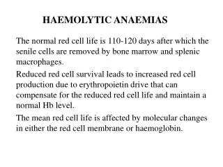
Normal red cell destruction Red cell destruction usually occurs after a mean lifespan of 120 days when the cells are removed extravascularly by the macrophages of the reticuloendothelial (RE) system, especially in the marrow but also in the liver and spleen.
Red cell metabolism gradually deteriorates as enzymes are degraded and the cells become non‐viable. The breakdown of haem from haemoglobin liberates iron for recirculation via plasma transferrin mainly to marrow erythroblasts, and protoporphyrin , which is broken down to bilirubin.
Bilirubin circulates to the liver where it is conjugated to glucuronides, which are excreted into the gut via bile and converted to stercobilinogen and stercobilin (excreted in faeces) Stercobilinogenand stercobilin are partly reabsorbed and excreted in urine as urobilinogenand urobilin..
Globin chains are broken down to amino acids which are reutilized for general protein synthesis in the body. Haptoglobinsare proteins in normal plasma which bind haemoglobin. The haemoglobin–haptoglobin complex is removed by the RE system.
Intravascular haemolysis (breakdown of red cells within blood vessels) plays little or no part in normal red cell destruction
Haemolysis Definitions Haemolysis indicates that the destruction of red cells is accelerated. Haemolyticanaemiasare defined as anaemias that result from an increase in the rate of red cell destruction.
Normally, in adults, the bone marrow output is well below its maximal capacity. Red cell production can be increased more than ten foldin the adult by increasing the cellularity of existing haemopoieticmarrow, as well as by expansion of haemopoietic marrow into the long bones.
In the newborn, and during infancy, marrow expansion depends on expanding the medullary cavity of bones, leading to thinning of cortical bone. These bony changes are most extreme in the β-thalassaemia syndromes, but some skeletal changes, usually some bossing of the frontal bones, may be seen in more extreme hereditary haemolyticanaemias of other causes.
Increased red cell destruction is often completely matched by increased production, resulting in Compensated haemolysis.
When the rate of haemolysis exceeds the maximum erythropoieticcapacity of the bone marrow, or when the latter is limited (e.g. because of inadequate supply of iron or folateor by ineffective erythropoiesis), the result is Haemolyticanaemia
Anaemiadue to haemolysis may not be seen until the red cell lifespan is less than 30 days.
Hereditary haemolyticanaemias are the result of ‘intrinsic’ red cell defects, whereas acquired haemolyticanaemias are usually the result of an ‘extracorpuscular’ or ‘environmental’ change. Paroxysmal nocturnal haemoglobinuriaPNH) is the exception because, although it is an acquired disorder, the PNH red cells have an intrinsic defect
General features of haemolysis The clinical and laboratory aspects of haemolysis depend on the consequences of increased red cell destruction and production as well as the main process by which destruction takes place..
In the presence of haemolysis, serum haptoglobin levels are greatly reduced or absent. However, haptoglobin is an acute-phase protein and levels will increase in the presence of inflammation. Haemopexin is another haem-binding protein produced by the liver that is decreased in haemolysis
Chronic haemolyticanaemiamay increase the iron content of the body through increased iron absorption as a result of anaemiacoupled to the retention of the haemiron following binding to haptoglobin and haemopexin. In rare cases of inherited haemolyticanaemia, this iron overload may be sufficient to produce clinically important effects, particularly if there is coinheritance of a haemochromatosis gene. .
In most haemolyticanaemias, owing to membrane defects, the destruction of red cells takes place extravascularly in the reticuloendothelial system, particularly in the spleen, and the iron is retained. When destruction is intravascular, free haemoglobinwill be released into the plasma, producing haemoglobinaemia and methaemalbuminaemia, and will pass through the glomerulus to produce haemoglobinuria and haemosiderinuria.
Iron deficiency is thus more likely than overload in intravascular haemolysis
Clinical features -pallor -Mild fluctuating jaundice and splenomegaly. -There is no bilirubin in urine but this may turn dark on standing because of excess urobilinogen. -Pigment (bilirubin) gallstones may complicate the condition . - Some patients (particularly with sickle cell disease) develop ulcers around the ankle . -.
Jaundice, anemia, and hemoglobinemia from intravascular hemolysis
-Aplastic crises may occur, usually precipitated by infection with parvovirus which ‘switches off’ erythropoiesis, and are characterized by a sudden increase in anaemia and drop in reticulocyte count .. -Rarely, folate deficiency may cause an aplastic crisis in which the bone marrow is megaloblastic
Laboratory findings 1. Features of increased red cell breakdown: (a)Raised serum unconjugated bilirubin. (b) urine urinobilinogen increased; (c) serum haptoglobins absent because the haptoglobins become saturated with haemoglobin and the complex is removed by RE cells. .
2 Features of increased red cell production: a.Marrowexpansion: bone changes b.Increasederythropoiesis: ↓ myeloid/erythroid ratio c.Reticulocytosis: polychromasia d.Increasedfolate requirements: macrocytosis.
Bony changes include bossing of the skull; hypertrophy of the maxilla, exposing the upper teeth; depression of nasal bridge; and periorbital puffiness
Bone marrow findings (upper :B.M.aspirate ,lower:B.M.biopsy) in hemolytic anemia. Erythroid hyperplasia is present with a predominance of erythroid precursors
3 Damaged red cells: (a) morphology (e.g. microspherocytes, elliptocytes, fragments); (b) osmotic fragility; (c) specific enzyme, protein or DNA tests
Warm autoimmune haemolyticanaemia. Blood filmshowing spherocytosis (arrows), polychromasia and a nucleatedred blood cell
Cold haemagglutinin disease. Blood film showing gross haemagglutination
Osmotic fragility curves of normal and hereditary spherocytosis red blood cells
Intravascular and extravascular Haemolysis Two mechanisms:
1-Extravascular haemolysis excessive removal of red cells by cells of the RE system . 2.Intravascular haemolysis RBC are broken down directly in the circulation
Whichever mechanism dominates will depend on the pathology involved.
The main laboratory features of intravascular haemolysis : 1 .Haemoglobinaemiaand haemoglobinuria. 2 .Haemosiderinuria. 3 .Methaemalbuminaemia(detected spectrophotometrically).
Causes of intravascular haemolysis. • Causes of intravascular haemolysis. -Mismatched blood transfusion (usually ABO) -G6PD deficiency with oxidant stress -Red cell fragmentation syndromes -Some severe autoimmune haemolyticanaemias -Some drug‐ and infection‐induced haemolyticanaemias -Paroxysmal nocturnal haemoglobinuria -March haemoglobinuria -Unstable haemoglobin
Features of extravascular haemolysis -jaundice. • gallstones • splenomegaly • raised reticulocytes, unconjugated bilirubin and absent haptoglobins.
Hereditary haemolyticanaemias Membrane defects -Hereditary spherocytosis : Hereditary spherocytosis (HS) is the most common hereditary haemolyticanaemia in Northern Europeans. -The inheritance is autosomal dominant with variable expression; rarely it may be autosomal recessive
Pathogenesis • Defects in the proteins involved in the vertical interactions between the membrane skeleton and the lipid bilayer of the red cell . • The marrow produces red cells of normal biconcave shape but these lose membrane and become increasingly spherical (loss of surface area relative to volume) as they circulate through the spleen and the rest of the RE system. • The loss of membrane may be caused by the release of parts of the lipid bilayer that are not supported by the skeleton. Ultimately, the spherocytes are unable to pass through the microcirculation, especially in the spleen, and die prematurely
Clinical features -Anaemia can present at any age from infancy to old age. -Jaundice is typically fluctuating. • Splenomegaly occurs in most patients. -Pigment gallstones are frequent . • Aplastic crises, usually precipitated by parvovirus infection, may cause a sudden increase in severity of anaemia.