Download
1 / 58
590 likes | 1.06k Views
HAEMOLYTIC ANAEMIAS. The normal red cell life is 110-120 days after which the senile cells are removed by bone marrow and splenic macrophages.

E N D
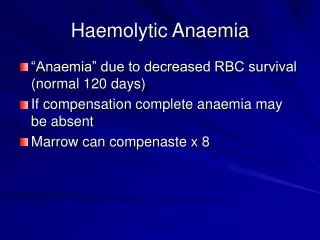
HAEMOLYTIC ANAEMIAS The normal red cell life is 110-120 days after which the senile cells are removed by bone marrow and splenic macrophages. Reduced red cell survival leads to increased red cell production due to erythropoietin drive that can compensate for the reduced red cell life and maintain a normal Hb level. The mean red cell life is affected by molecular changes in either the red cell membrane or haemoglobin.
A haemolytic state exists when the in vivo survival of the RBC is shortened. Anaemia occurs if the onset of haemolysis is sudden with no time for marrow compensation or in severe chronic haemolysis when the mean red cell life is very short.
CLINICAL FEATURES Jaundice: generally mild and often not noticed by the patient. Anaemia: recent onset = acquired long-standing = possibly congenital. Haemoglobinuria: intravascular haemolysis. Urobilinogenuria: increased Hb catabolism. Splenic pain: spenomegaly or splenic infarction. Leg ulcers: intrinsic red cell disorders, e.g. sickle cell disease. Skeletal hypertrophy: severe congenital haemolytic anaemias and thalassaemias.
CLASSIFICATION OF HAEMOLYTIC ANAEMIAS GENETIC EXAMPLES Membrane: Hereditary spherocytosis elliptocytosis, pyropoikilocytosis Haemoglobin: Haemoglobinopathies Unstable haemoglobins Thalassaemias Enzymes: G6PD or PK deficiency Other: Abeta-lipoproteinaemia
ACQUIRED EXAMPLES Immune: Isoimmune, autoimmune or drug Mechanical: March haemoglobinuria, burns or microangiopathy Infections: Malaria, Clostridium welchi. Bartonellosis (Oroya fever) Toxins: Arsenic, copper, lead, venoms Dyserythropoietic: PNH Other: Osmotic effects, liver disease, hypophosphataemia
LABORATORY FINDINGS Features of increased erythrocyte breakdown: • Unconjugated bilirubinaemia. • Urobilinogenuria. • Haptoglobins decreased. • Radioisotope red cell survival studies can quantitate rate and site of destruction.
Features of increased erythrocyte production: • Reticulocytosis • Polychromasia and nucleated red cells in peripheral blood film. • Erythroid hyperplasia in bone marrow aspirate. • Radiological changes, e.g. "hair on end" appearance of cranial X-ray.
Features specific to intravascular haemolysis: • Haemoglobinaemia • (haptoglobin and haemopexin exhausted). • Methaemoglobinaemia. • Haemoglobinuria. • Haemosiderinuria.
CONGENITAL HAEMOLYTIC ANAEMIAS Hereditary Spherocytosis (HS) Elliptocytosis (HE) Stomatocytosis (Hydrocytosis) Pyropoikilocytosis (HPP)
MEMBRANE DISORDERS Hereditary spherocytosis (HS): Inheritance mostly autosomal dominant transmission. There is a subnormal level of spectrin in the red cell cytoskeleton. 25% of cases have little evidence of haemolysis or splenomegaly unless stressed (infection or pregnancy). 60% have chronic anaemia with variable jaundice and splenomegaly. Ultrasound examination often shows gallstones from increased bilirubin excretion. Rarely patients present with transfusion-dependent anaemia.
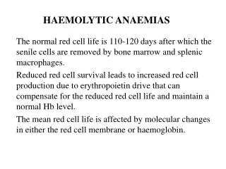
Spherocytes have lost surface area relative to volume and show decreased ability to withstand osmotic stress. They are most vulnerable in the spleen (most patients respond well to splenectomy). Not all the cells are spheres; the osmotic fragility curve may just have a "tail" where osmotic lysis is increased. The AHG (Coomb's) test is negative. Retics are 5-25%. The MCHC is often raised. ATP and glucose utilisation are increased to fuel the cation pumps which transport sodium out of the cell (HS cells accumulate sodium). Autohaemolysis gives a Type I result (added glucose reduces haemolysis). Family studies are essential.
100 80 60 40 20 Sickle Cell Disease -Thalassaemia Major Hereditary Spherocytosis Auto-immune HA Normal range % Lysis 1 2 3 4 5 6 7 8 9 NaCl g/L Osmotic Fragility
Hereditary elliptocytosis (HE): an autosomal dominant gene transmits both benign HE and haemolytic HE. A skeletal protein deficiency or defect in the red cell membrane exists. Spectrin or associated proteins (band 4.1 and ankyrin) may be involved. Benign HE presents with variable red cell shape (from ovalocytes to rods) and osmotic fragility. In haemolytic HE schistocytes and spherocytes are often seen. Splenectomy is rarely necessary.
HEREDITARY STOMATOCYTOSIS (Hydrocytosis): Autosomal dominant transmission. 10-40% of cells show slits and there may be mild haemolysis. The red cell cation (Na) content is increased with resultant influx of water and increased osmotic fragility. The MCV is high.
HEREDITARY PYROPOIKILOCYTOSIS (HPP): Autosomal recessive inheritance. Patients present with micropoikilocytosis, schistocytes, microspherocytes and elliptocytes. The MCV is very low (55-65 fl). The red cells fragment in vitro at 450C; normal cells fragment at 490C. This phenomenon is due to thermal instability of spectrin within the cytoskeleton. HPP is actually a subgroup of common HE.
DEFECTS OF RED CELL METABOLISM Defects of red cell metabolism should be suspected in any case of non-spherocytic haemolytic anaemia where there is no obvious acquired cause.
Pentose Phosphate Pathway (PPP) enzymopathies are commoner than those of the Embden-Meyerhof Pathway (EMP) but their clinical effects are less severe. The PPP is responsible for generating reducing power in the form of reduced glutathione and NADPH that counteract oxidant stresses. In contrast to EMP enzyme defects, haemolysis is increased by oxidant drug ingestion. Most PPP enzyme deficiencies lead to the production of Heinz bodies. These red cell inclusions stain with methyl violet. They represent insoluble aggregates of partially denatured haemoglobin. Bite cells result from the removal (pitting) of Heinz bodies by macrophages.
G-6-PD DEFICIENCY The gene for G6PD is carried on the X chromosome and so is expressed by hemizygous males and homozygous females. Over 300 variants of the enzyme have been described. Electrophoresis shows a fast moving form GdA, and a slower-moving form, GdB.
Most G6PD deficients are asymptomatic but there is great variability in susceptibility. 7-15% of African Negroes have a defective form called GdA-. Young cells show normal activity but the enzyme is unstable and cells become deficient on ageing. This African type of G6PD deficiency is milder than the Mediterranean type which cells of all ages are affected. Other forms include the Canton and Japanese varieties. G6PD deficient red cells are resistant to P. falciparum malaria parasites.
Agents causing haemolysis in G6PD deficient subjects include: antimalarials antipyretics sulphonamides ascorbic acid vitamin K naphthalene (used in mothballs) Favism refers to haemolysis initiated by eating the Vicia fava (broad) bean or by inhaling its pollen.
LABORATORY TESTS FOR G6PD DEFICIENCY G6PD deficiency in male or homozygous female patients can be detected by screening tests but it is more difficult to detect heterozygous (female) carriers. The enzyme activity depends on red cell age with the highest activity in reticulocytes. Testing should be performed when any reticulocytosis has abated. Retics can be physically removed by centrifuging the whole blood specimen and sampling from the bottom of the red cell mass. This applies to any specimen, including neonatal, with retics >5%.
The G6PD fluorescent spot screening test is based upon the production of NADPH which fluoresces under longwave UV light. • The test is excellent for low level hemizygotes but heterozygotes may not be detected. • G6PD • G6P + NADP ----------> 6 Phosphogluconate + NADPH • (no fluorescence) (fluorescence)
Heinz Body Haemolytic Anaemia Heinz bodies G6PD deficiency is the most common enzymopathy causing hereditary haemolytic anaemia. Bite cells and helmet cells Blister cell
Embden-Meyerhof Pathway Enzymopathies These are mostly autosomal recessive disorders producing congenital non-spherocytic haemolytic anaemias of variable severity. Morphological features include finding prickle cells. Pyruvate kinase (PK) deficiency is the commonest of the glycolytic pathway enzymopathies. Pyruvate kinase deficiency is inherited as an autosomal recessive trait affecting both sexes equally.
PK catalyses the conversion of phosphoenolpyruvate to pyruvate with the production of ATP. PK-deficient red cells are deficient in ATP and their lifespan is thus shortened. Retics have an alternative source of ATP via Krebs cycle and are usually increased in PK deficient subjects. Jaundice, splenomegaly and haemolysis may be the presenting features in infancy but many cases are not detected until adult life. Owing to the position of PK in the EMP there is a build up of 2,3-DPG with resultant reduction in oxygen affinity and so the anaemia is relatively well tolerated.
Diagnosis rests on the demonstration of reduced PK activity while increased levels of 2,3-DPG provides supportive evidence. Autohaemolysis is Type II (haemolysis is not reduced with the addition of glucose).
ACQUIRED HAEMOLYTIC ANAEMIAS Immune mediated haemolysis is caused by the attachment of antibody to the red cell membrane. The rate and site of haemolysis depends on the type of antibody (IgG or IgM) and its ability to fix complement.
The optimal temperature for binding of antibody is important. Usually, IgG antibodies are warm (370C) and IgM are cold reacting. Cells coated with IgG are removed directly by splenic macrophages that bear Fc receptors. Both IgG and IgM can fix complement but usually only part of the sequence is fixed, thus avoiding intravascular haemolysis. Cells coated with C3 are removed by macrophages of the RE system but this component has a short half-life and gives rise to C3d to which macrophage receptors are insensitive.
Part of the red cell membrane is lost during immune adherence and gives rise to spherocytes that are a feature of warm autoimmune haemolytic anaemia (AIHA). Cold-reacting IgM antibodies are more likely to trigger the entire complement cascade and give rise to intravascular haemolysis.
Classification Of Immune Haemolytic Anaemias 1. Alloimmune: Transfusion reactions, Haemolytic Disease of the Newborn (HDN) 2. Autoimmune: Warm AIHA, Cold AIHA, Paroxysmal Cold Haemoglobinuria (PCH) 3. Drug-induced: Immune complex, drug adsorption (hapten), methyldopa-induced
ANTIGLOBULIN TESTS The direct antiglobulin test (DAT) is designed to react with cells bearing IgG and/or complement. IgM always binds complement so the reagent does not contain anti-IgM. Washed red cells are mixed directly with the antiglobulin reagent and agglutination recorded. The DAT is positive in most cases of AIHA. Monospecific reagents demonstrate IgG alone or together with C3 components. In immune haemolysis associated with SLE, IgG is detected together with C3. Only C3 is present in cold AIHA.
The indirect antiglobulin test (IAT) is used to detect free antibody in the patient's serum using cells with known antigenic characteristics. It is often positive in severe AIHA. An eluate of antibody from the patient's cells can be used to determine antigenic specificity.
Cold haemagglutinin disorders are characterised by in vivo agglutination of red cells at temperatures below 370C. • Cold autoantibodies are responsible for: • secondary cold AIHA • cold haemagglutinin disease (CHAD) • paroxysmal cold haemoglobinuria (PCH)
50% of patients infected with Mycoplasma pneumoniae show a rise in the titre of anti-I, an IgM cold antibody normally present in low titre. This antibody reacts optimally with adult red cells in the cold. Neonatal red cells only express i antigen and are therefore not agglutinated. Following infectious mononucleosis (IM) a similar cold-reacting antibody, anti-i, is frequently produced which in rare cases can affect adult red cells. Cold antibodies produced secondary to these infections are polyclonal (contain both lambda and kappa light-chains).
The anti-I found in cold haemagglutinin disease (CHAD) and cold haemagglutination secondary to lymphomas is a monoclonal (IgM with kappa light-chain specificity). The low thermal amplitude of these antibodies results in red cell agglutination in the cooler peripheral circulation of the hands, feet, nose and ears. Intravascular haemolysis occurs if sufficient complement is bound.
Paroxysmal Cold Haemoglobinuria PCH is characterised by haemoglobinuria following cold exposure. It occurs in children in association with viral disorders and is caused by the Donath-Landsteiner antibody, an IgG biphasic haemolysin with anti-P specificity. Diagnosis rests on the demonstration of in vitro haemolysis when red cells and serum are first chilled (00C) and then rewarmed (370C). PCH is an acute haemolytic anaemia occurring almost exclusively in children and young adults.
DRUG INDUCED HAEMOLYTIC ANAEMIAS • Immune Complex Mechanism • Drug Adsorption Mechanism • Methyldopa-induced Mechanism
Immune Complex Mechanism (acute intravascular haemolysis) The drug, e.g. quinidine, provokes antibody formation and the resulting immune complex is loosely adsorbed onto the red cell where it can fix complement, detectable by the DAT. The red cells become "innocent bystanders".
Drug Adsorption Mechanism (slow reacting). The drug, e.g. penicillin, binds to the red cell and IgG against the drug then attaches leading to extravascular destruction of coated cells. The drug may act as a hapten and only provoke antibody formation after attachment to the red cell or after reacting with certain serum proteins. The DAT is positive due to IgG sensitisation. Intravascular haemolysis can occur if complement is fixed.
Methyldopa-induced Mechanism. This was first reported in association with methyldopa. L-dopa and aldomet can cause haemolysis by a similar mechanism. After at least four months of treatment, 15-20% of patients develop a positive DAT due to IgG coating but less than 1% show evidence of haemolysis. The antibody often shows Rh specificity (anti-e). Haemolysis is extravascular.
NON-IMMUNE HAEMOLYTIC ANAEMIA • Haemolysis Caused Directly by Infection • Mechanical Causes of Haemolysis • Chemical and Physical Causes of Haemolysis • Acquired Membrane Disorders
Haemolysis Caused Directly by Infection Intracellular infection: Malaria and Babesiosis (parasites physically disrupt the red cell membrane). Extracellular Infection: Bartonella and Clostridia welchi attack the red cell membrane with enzymes. Bacterial sepsis produces haemolysis secondary to DIC. Red cells are also vulnerable to certain viruses.
MECHANICAL CAUSES OF HAEMOLYSIS Red cells are flexible and must deform many times during their lifespan to pass through small capillaries. Fragmentation occurs when excessive shear stresses are applied resulting in intravascular haemolysis and/or the formation of red cell fragments (schistocytes).
Cardiac Prosthesis Shear stresses are caused by turbulent blood flow in a high pressure system. This arises around prosthetic heart valves, particularly if the valve malfunctions.
Micro-Angiopathic Haemolytic Anaemia (MAHA) results from small blood vessel abnormalities that impede blood flow, e.g. fibrin deposition, microthrombi and disseminated intravascular coagulation (DIC). The coagulation screen may be abnormal with associated thrombocytopenia due to chronic DIC.
Thrombotic Thrombocytopenic Purpura (TTP) is related to aggregation of platelets in small capillaries with consequent occlusion of the vessel by platelet plugs and fibrin. Haemolytic Uraemic Syndrome (HUS); there is endothelial damage with fibrin deposition in small vessels. HUS is characterised by intravascular haemolysis, renal failure and thrombocytopenia.
March Haemoglobinuria First described in a German soldier after marching exercise. The anaemia has been described in joggers and marathon runners. Red cells are disrupted as they pass through capillaries subjected to pounding. Haemoglobinuria is only present following exercise.
A careful history and examination will often reveal the cause of mechanical haemolysis as indicated by the presence of red cell fragments in the peripheral blood. Perls's stain of urinary sediment can show the presence of iron; a useful test for any chronic low grade intravascular haemolysis when there is little evidence on blood film inspection.
CHEMICAL AND PHYSICAL CAUSES OF HAEMOLYSIS • Oxidative Haemolysis • Nonoxidative Haemolysis • Osmotic Effects • Burns
Oxidative Haemolysis: Oxidative stress on the red cell may affect the haemoglobin molecule with either the globin chains or the haem group being affected. Most oxidizing agents will produce either Heinz body formation by denaturing globin chains or by oxidizing the haem group to produce methaemoglobinaemia.